What is a Terrible Triad Injury of the Elbow?
A Terrible Triad Injury of the Elbow is a severe and complex injury that involves:
- Elbow Dislocation: The elbow bones are forced out of their normal positions.
- Radial Head or Neck Fracture: The radial head or neck, part of the radius bone near the elbow, is fractured.
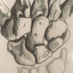
- Coronoid Process Fracture: The coronoid process is fractured, a triangular part of the ulna bone.
Diagnosis
- X-rays: The primary tool for diagnosing the injury. They help visualise the position of the bones and identify fractures.
- CT Scans: These are often used to get a more detailed view of the fractures, especially useful for surgical planning.
Treatment Options
- Surgical Treatment:
- Open Reduction and Internal Fixation (ORIF): Surgical method to fix the fractures using plates, screws, or rods.
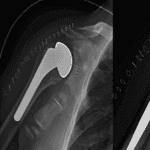
- Radial Head Arthroplasty: Replacement of the radial head with a prosthetic implant.
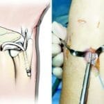
- Lateral Collateral Ligament (LCL) Reconstruction: Repairing the ligament on the outer side of the elbow.
- Coronoid ORIF: Surgical fixation of the coronoid fracture.
- Medial Collateral Ligament (MCL) Reconstruction: Repairing the ligament on the inner side of the elbow if necessary.
Causes and Mechanism
- Causes: Typically occurs from a high-energy impact, such as falling onto an outstretched hand with the arm extended.
- Mechanism:
- The elbow is subjected to valgus (outward), axial (along the arm’s length), and posterolateral rotatory (twisting) forces.
- This combination leads to dislocation and fractures, starting from the lateral side and moving medially.
- Pathoanatomy:
- The LCL is the first structure to fail.
- The anterior capsule of the elbow is injured next.
- MCL disruption can occur if the force is significant enough.
Anatomy Involved
- Radial Head:
- Key to preventing posterolateral rotatory instability (PLRI).
- Acts as a secondary stabilizer against valgus forces.
- Coronoid Process:
- Provides stability to the ulnohumeral joint.
- Prevents posterior subluxation (partial dislocation) beyond 30 degrees of flexion.
- The fracture often includes part of the anterior capsule, which aids in repair.
- Medial Collateral Ligament (MCL):
- Composed of three parts: anterior bundle (most important for stability), posterior bundle, and transverse ligament.
- Essential for resisting valgus and posteromedial rotatory forces.
- Lateral Collateral Ligament (LCL):
- Comprises four parts: lateral ulnar collateral ligament (LUCL), radial collateral ligament (RCL), annular ligament, and accessory collateral ligament.
- Crucial for preventing posterolateral rotatory instability.
- Often avulsed (torn off) from the lateral epicondyle during injury.
Symptoms and Examination
- Symptoms:
- Severe pain in the elbow.
- Clicking and locking sensation, especially when extending the elbow.
- Physical Exam:
- May reveal instability patterns (varus or valgus).
- The distal radial ulnar joint should be checked for Essex-Lopresti injury (a severe injury involving the radial head and the interosseous membrane).
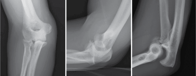
Imaging
- Radiographs (X-rays):
- Essential for evaluating the alignment of the ulnohumeral and radiocapitellar joints.
- Look for coronoid fractures.
- Both pre-reduction (before the bones are set) and post-reduction films are needed.
- Additional wrist and forearm X-rays may be necessary.
- CT Scans:
- Provide a detailed view of the coronoid fracture.
- 3D imaging can help determine the exact nature of the fracture lines.
Treatment Details
Nonoperative Treatment
- Immobilization:
- The elbow is immobilized at 90 degrees of flexion for 7-10 days.
- Indicated in rare cases where the joints are stable, fractures are small, and early range of motion is feasible.
- Technique:
- After a week of immobilization, gradual progression to active motion begins.
- Initial active motion with the elbow at 90 degrees and forearm pronated, avoiding terminal extension.
- Static progressive extension splinting at night after 4-6 weeks.
- Strengthening exercises start after six weeks.
Operative Treatment
- Indications:
- Required for unstable injuries with significant fractures and dislocations.
- Techniques:
- ORIF or Radial Head Arthroplasty: Depending on the fracture type and severity.
- LCL Reconstruction: Using suture anchors or transosseous sutures.
- MCL Reconstruction: If instability persists after addressing other structures.
- Surgical Approach:
- A posterior skin incision provides access to the elbow’s medial and lateral aspects.
- This approach is more cosmetic and has a lower risk of injuring cutaneous nerves.
- Specific Techniques:
- Radial Head ORIF: Using screws and plates if fractures are less than 40% of the articular surface.
- Radial Head Arthroplasty: For comminuted fractures (more than three pieces).
- Coronoid ORIF: Using sutures, suture anchors, screws, or plates.
- LCL Repair: Reattachment with sutures or anchors at the lateral epicondyle.
- MCL Repair: Indicated for persistent instability after other repairs.
Complications
- Instability: More common with Type I or II coronoid fractures.
- Failure of Fixation: Often seen in radial neck fractures due to poor vascular supply.
- Stiffness: Very common; early ROM exercises are crucial.
- Heterotopic Ossification: Prophylaxis may be necessary in high-risk patients.
- Post-Traumatic Arthritis Can result from initial cartilage damage or residual instability.
Prognosis
- Historically, outcomes have been poor due to:
- Persistent instability.
- Joint stiffness.
- Development of arthritis.
- Early and appropriate treatment is essential for better outcomes and to minimise complications.
Videos :