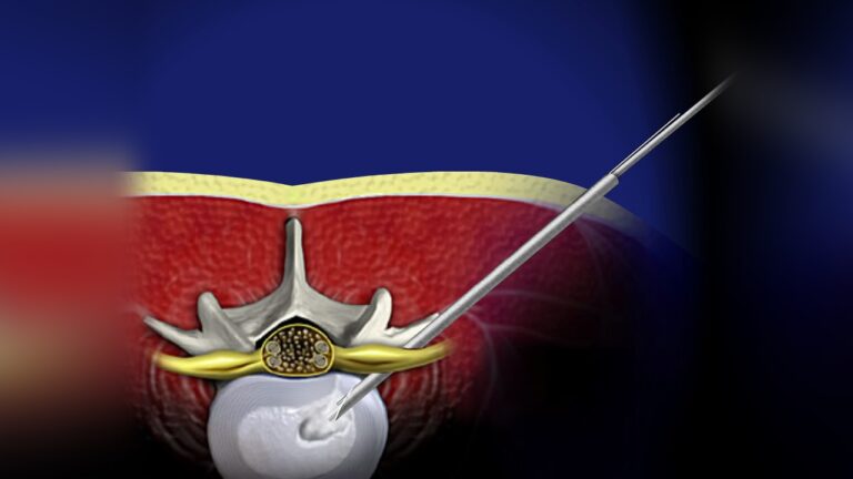
Endoscopic Diskectomy

Overview of Endoscopic Diskectomy

Endoscopic spine surgery is an advanced, minimally invasive procedure used to treat herniated disks in the lower back. This technique involves the use of an endoscope—a thin, flexible tube equipped with a camera and light source—that allows surgeons to access and visualize the spine through small incisions.

With its numerous potential benefits, endoscopic diskectomy has become a popular and effective alternative to traditional open spine surgery and microdiskectomy, offering faster recovery and reduced tissue damage.

Description of a Herniated Disk

A disk herniates when its jelly-like nucleus begins to push against the outer ring due to wear and tear or a sudden injury. This pressure on the outer ring can cause lower back pain.

If the pressure persists, the nucleus may fully push through the outer ring or cause it to bulge. This can exert pressure on the spinal cord and nerve roots, leading to both mechanical compression and chemical irritation from the disk material.

When the nerve roots become inflamed or compressed, symptoms such as:

  • Pain,
  • Numbness, and
  • Weakness in one or both legs may occur—a condition known as sciatica.

Endoscopic diskectomy aims to relieve these symptoms by removing the herniated portion of the disk while preserving surrounding tissues.
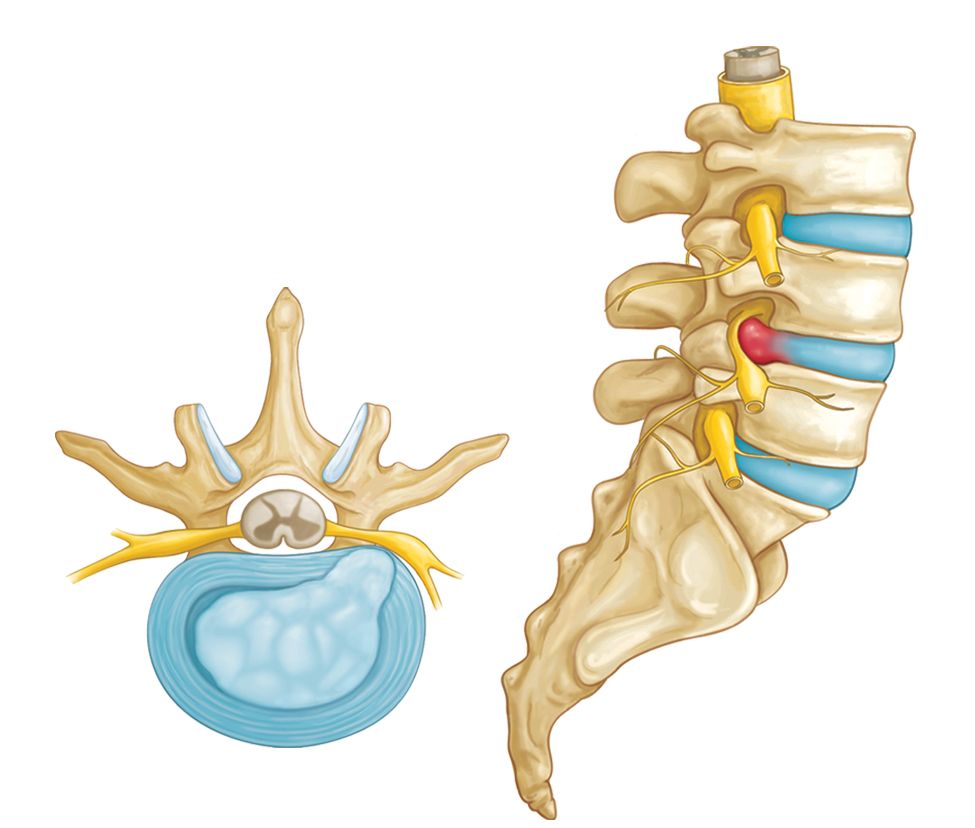
In a herniated disk, the soft, jelly-like center of the disk can push all the way through the outer ring.  (Side and cross-section views shown.)

Lumbar Diskectomy: When is Surgery Recommended?

Lumbar diskectomy, a surgical procedure to treat herniated disks in the lower back, is not necessary for most patients. Spine surgery is typically recommended only when nonsurgical treatments fail to alleviate symptoms or in cases involving severe or specific conditions, such as:

  • Persistent Muscle Weakness: Ongoing weakness that interferes with daily activities.
  • Difficulty Walking: Pain, weakness, or reduced mobility that impacts the ability to walk.
  • Loss of Bladder or Bowel Control: A serious sign of severe nerve compression that requires immediate medical attention.

The decision to proceed with lumbar diskectomy is made after careful consideration of the patient’s symptoms, overall health, and lifestyle.


Evolution of Diskectomy Techniques

Historically, herniated disks were removed using a large open incision. Advances in surgical technology have led to minimally invasive procedures like microdiskectomy and endoscopic diskectomy, which offer significant benefits.

Microdiskectomy

  • A commonly used minimally invasive procedure.
  • Performed using a microscope to relieve pressure on the spinal nerve.
  • Aims to reduce pain and improve mobility with minimal tissue disruption.

Endoscopic Diskectomy

  • A newer technique involving an endoscope, a thin tube with a camera, to visualize and access the affected area.
  • The minimally invasive approach reduces incision size, recovery time, and surgical trauma.
  • Comparable to arthroscopic techniques used in knee, hip, and shoulder surgeries, making it a highly effective alternative to traditional methods.

Both techniques aim to relieve nerve compression and associated symptoms, improving patients’ quality of life with quicker recovery times.

Potential benefits of endoscopic diskectomy include:

  • Smaller incisions
  • Reduced scarring
  • A shorter recovery time

In cases where there are disk herniations at multiple levels of the spine, an open procedure with a larger incision may be necessary.

Procedure: What Happens During an Endoscopic Diskectomy?

During an endoscopic diskectomy, the following steps are performed to treat a herniated disk:

  1. Anesthesia:
    • The patient is placed under general anesthesia (asleep during the procedure) or given local anesthesia with sedation (awake but relaxed, with the treatment area numbed).
  2. Small Incisions:
    • The surgeon makes either a single 1 cm incision or two smaller 7 mm incisions to access the spine near the herniated disk.
  3. Guide Wire Placement:
    • A guide wire is inserted through the incision and carefully directed toward the affected disk using fluoroscopic (X-ray) guidance.
  4. Endoscope Insertion:
    • The surgeon threads an endoscope (a thin tube with a camera) over the guide wire to the area of the herniated disk.
    • The endoscope provides a clear view of the disk and surrounding structures on a video monitor. Water is used to enhance visualization, offering greater magnification of the spine.
  5. Disk Removal:
    • Specialized instruments are passed through the endoscope to remove the herniated portion of the disk or any fragments pressing on the spinal nerves.
  6. Closure:
    • After the procedure, the surgeon removes the endoscope and closes the incision(s) with sutures or adhesive strips.

This minimally invasive approach allows for precise treatment with minimal disruption to surrounding tissues.

Advantages and Disadvantages of Endoscopic Diskectomy

Advantages

Endoscopic diskectomy offers several potential benefits compared to traditional open surgery:

  • Smaller Incisions: Minimizes tissue damage, leading to less pain after surgery.
  • Minimal Scarring: Results in a more aesthetically pleasing appearance post-surgery.
  • Shorter Hospital Stay: Many patients undergo this as an outpatient procedure, returning home the same day.
  • Faster Recovery: Patients can resume daily activities sooner.
  • Lower Risk of Complications: Reduces the likelihood of infections, excessive bleeding, and other complications.

Disadvantages

Despite its benefits, endoscopic diskectomy also has limitations:

  • Specialized Training: Requires expertise, which limits the number of experienced surgeons available.
  • Limited Suitability: Not appropriate for all types of herniated disks, especially complex or multi-level herniations.
  • Learning Curve: Surgeons new to the technique may have higher rates of complications or incomplete removal of the herniated disk.
  • Complications: Risks include infection, bleeding, nerve damage, or dural tears (tears in the tissue protecting the spinal cord). In some cases, symptoms may persist or return after surgery.
  • Higher Cost: The procedure is more expensive due to specialized equipment and expertise.
  • Long-Term Outcomes: More research is needed to compare its durability and effectiveness to traditional open surgery.

Recovery After Endoscopic Diskectomy

The recovery process for endoscopic diskectomy is typically faster than that of open surgery:

  • Outpatient Procedure: Most patients go home the same day or after a short hospital stay.
  • Pain Management: Post-operative discomfort is usually manageable with medication.
  • Activity Restrictions: Physical activity may be limited for a few weeks, and physical therapy is often recommended to regain strength and mobility.
  • Gradual Return to Normalcy: Patients are encouraged to ease back into their daily routines over time.
  • Follow-Up Appointments: Regular follow-ups are scheduled to monitor progress and address any complications.

Future Directions of Endoscopic Diskectomy

As this technique evolves, its potential continues to grow:

  1. Refinement of Techniques: Ongoing improvements in surgical instruments and methods will make the procedure more precise and effective.
  2. Enhanced Surgeon Training: Comprehensive training programs will help more surgeons develop the expertise required, reducing associated risks.
  3. Long-Term Studies: Research on the durability and effectiveness of endoscopic diskectomy compared to traditional open surgery will help define its role in spinal care.

Endoscopic diskectomy shows promise as a minimally invasive solution, and advancements in technology and training are likely to enhance its outcomes and accessibility in the future.

Scroll to Top