Understanding Hip Dysplasia in Adolescents: Causes, Symptoms, and Treatment Options

The hip is a ball-and-socket joint, where the rounded top of the femur (thighbone) fits securely within a curved socket in the pelvis known as the acetabulum. In adolescents with hip dysplasia, however, the joint has not formed as it should—the acetabulum is too shallow to fully support and cover the femoral head. This structural irregularity can lead to hip pain and may accelerate the onset of osteoarthritis, a degenerative joint condition where the protective cartilage wears down, causing bone-on-bone contact.

Adolescent hip dysplasia often originates from developmental dysplasia of the hip (DDH), a condition present at birth or in early childhood. Although newborns are regularly screened for DDH, some cases remain unnoticed or are mild enough to go untreated. As a result, symptoms of hip dysplasia may not manifest until adolescence.

The primary goal in treating adolescent hip dysplasia is to alleviate pain and maintain the natural hip joint for as long as possible. Often, this involves surgical intervention to correct the joint’s anatomy, potentially delaying or preventing the progression of painful osteoarthritis.

Anatomy

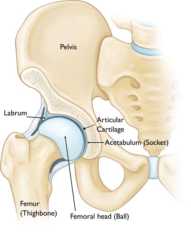
The hip is among the largest joints in the human body, functioning as a ball-and-socket structure. The “socket” component, known as the acetabulum, is part of the large pelvic bone. The “ball” is formed by the femoral head, which is the upper portion of the femur (thighbone). Both the femoral head and the acetabulum are coated with articular cartilage—a smooth, slippery tissue that cushions the bones, allowing them to move fluidly and reducing friction.

Surrounding the acetabulum is a ring of durable fibrocartilage called the labrum. This labrum acts like a gasket, creating a snug seal around the socket and stabilizing the femoral head within the joint.


The hip is a ball-and-socket joint. In a healthy hip, the head of the femur (ball) stays firmly within the acetabulum (socket).


Description

In individuals with hip dysplasia, the acetabulum is underdeveloped, making it too shallow for the femoral head to fit securely within the socket.

This anatomical irregularity disrupts the typical distribution of forces across the joint, causing the labrum to absorb more stress than usual. Consequently, excessive force concentrates on a smaller area of the hip cartilage and labrum. Over time, this can lead to the gradual wearing away of the articular cartilage, while the labrum may become torn or damaged. Such degenerative changes heighten the risk of developing early-onset osteoarthritis.

The extent of hip dysplasia can vary significantly:

  • In mild cases, the femoral head may simply have some looseness within the socket.
  • In more severe instances, the joint may exhibit complete instability, or the femoral head may be fully dislocated from the socket.

Cause

Adolescent hip dysplasia often stems from developmental dysplasia of the hip (DDH), a condition that may go unnoticed or untreated during infancy or early childhood.

DDH can run in families, passed down from one generation to the next. It can affect either hip but typically involves the left hip. While it can develop in anyone, certain factors increase the likelihood:

  • Female gender
  • Firstborn status
  • Breech birth position (bottom-first instead of head-first)

Symptoms

Hip dysplasia in young children generally does not cause pain. However, as time passes, altered forces within the hip joint lead to degenerative changes—gradual damage to the articular cartilage and labrum—which can become painful.

Commonly, the pain associated with hip dysplasia:

  • Centers in the groin area, though it may occasionally radiate to the outside of the hip
  • Starts as mild and intermittent but can become more frequent and intense over time
  • Worsens with activity or toward the end of the day

Some individuals may also experience sensations of locking, catching, or popping in the groin.

Doctor Examination

Physical Examination

During a physical exam, the doctor will review your child’s medical history and discuss symptoms. They will move the hip in various directions to evaluate the range of motion and attempt to replicate any pain or discomfort.

Imaging Tests

In many cases, a physical examination is sufficient to diagnose adolescent hip dysplasia. However, doctors may recommend imaging tests to exclude other potential causes of pain and to confirm the diagnosis.

  • X-rays: X-rays provide images of bones and help the doctor assess the alignment between the acetabulum and femoral head. They can also reveal signs of arthritis.
  • Computerized Tomography (CT) Scans: More detailed than standard X-rays, CT scans offer a clearer view of the severity of dysplasia.
  • Magnetic Resonance Imaging (MRI) Scans: MRIs create detailed images of soft tissues, which can help the doctor detect damage to the labrum and articular cartilage.


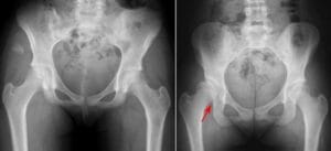
(Left) This X-ray shows two normal hips. (Right) This X-ray shows a dysplastic hip (arrow). The hip socket is shallow and there is only partial coverage of the femoral head.


Treatment

The primary goal in treating adolescent hip dysplasia is to delay or prevent osteoarthritis while preserving the natural hip joint for as long as possible.

Nonsurgical Treatment

If the dysplasia is mild and there is no damage to the labrum or articular cartilage, nonsurgical treatment may be recommended. Additionally, nonsurgical approaches may be considered initially for cases where joint damage is so extensive that a total hip replacement is the only surgical alternative.

Common nonsurgical treatments for adolescent hip dysplasia include:

  • Observation: For mild dysplasia with minimal symptoms, the doctor may recommend regular monitoring to track any progression. Follow-up appointments every 6 to 12 months will help ensure that any increase in pain, symptoms, or changes on X-rays are identified promptly.
  • Lifestyle Modifications: Your doctor might suggest avoiding activities that exacerbate pain. For adolescents who are overweight, weight loss can also help alleviate pressure on the hip joint.
  • Physical Therapy: Tailored exercises can improve hip mobility and strengthen the surrounding muscles, which can reduce strain on the labrum and cartilage.
  • Medications: Nonsteroidal anti-inflammatory drugs (NSAIDs), such as ibuprofen or naproxen, may relieve pain and reduce inflammation in affected joints.
  • Cortisone Injections: Cortisone, a potent anti-inflammatory medication, can be injected directly into the hip joint to temporarily relieve pain and inflammation.

Surgical Treatment

Surgery may be recommended if the adolescent experiences pain and has limited cartilage damage in the hip.

The most common surgical approach for treating hip dysplasia is an osteotomy, a procedure that involves reshaping and realigning the acetabulum or femur to improve joint positioning.

Various osteotomy techniques are available, and the specific type chosen depends on several factors, such as:

  • The adolescent’s age
  • Severity of dysplasia
  • Level of labrum damage
  • Presence of osteoarthritis
  • Years remaining for growth

Periacetabular Osteotomy (PAO): The most frequently performed osteotomy for adolescent hip dysplasia is the periacetabular osteotomy (PAO). “Periacetabular” refers to “around the acetabulum,” indicating that the procedure focuses on adjusting the acetabular region.


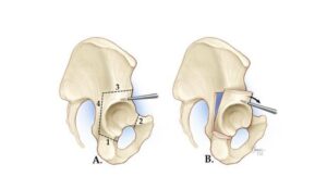
(A) In a periacetabular osteotomy, four cuts are made in the pelvic bone. (B) The surgeon uses a specialized tool to manipulate the bone fragment to gain access to the acetabulum.


In most cases, a periacetabular osteotomy (PAO) procedure takes between 2 to 3 hours. During this surgery:

  • The surgeon makes four strategic cuts around the pelvic bone to release the acetabulum.
  • The acetabulum is then rotated and repositioned to provide better coverage over the femoral head. X-rays are used throughout the procedure to guide the cuts and confirm the acetabulum’s new, optimal positioning.
  • Once the repositioning is complete, the surgeon secures the bone with screws to keep it stable as it heals.

Arthroscopy

In addition to PAO, hip arthroscopy may be performed to address a torn labrum.

During hip arthroscopy, the surgeon inserts a tiny camera, known as an arthroscope, into the hip joint. This camera projects images onto a monitor, allowing the surgeon to guide delicate surgical instruments. Common arthroscopic procedures include:

  • Labral Refixation: Here, the surgeon trims damaged tissue along the acetabular rim and reattaches the torn labrum to the bone for improved stability.
  • Debridement: In cases where labral tissue is torn or weakened, trimming the damaged portions can often alleviate pain.


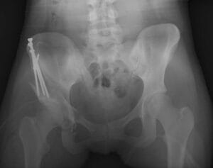
In this X-ray image, the acetabulum has been repositioned using a periacetabular osteotomy, and the bones are held in place with screws.


Complications

As with any surgery, periacetabular osteotomy (PAO) carries certain risks. Your surgeon will discuss each potential risk with you in detail and take specific precautions to minimize complications.

Although complications are uncommon, the most frequent ones include:

  • Infection
  • Blood clots
  • Injury to blood vessels or nerves
  • Persistent hip pain
  • Nonunion, or failure of the osteotomy site to heal

Recovery

After surgery, your child will stay in the hospital for 2 to 4 days to ensure proper monitoring and pain management.

Typically, full weight-bearing on the operated leg is restricted for 6 to 12 weeks to allow the bones to heal in their new position. During this period, your child will need to use crutches for mobility.

Around six weeks post-surgery, a follow-up visit with the doctor will include X-rays to assess healing progress. Based on the results, the doctor will advise when it’s safe to start weight-bearing on the leg and begin physical therapy. The physical therapist will guide your child through specific exercises aimed at maintaining range of motion and gradually restoring strength and flexibility in the hip joint.

Outcomes

Periacetabular osteotomy is generally effective in postponing the need for a hip replacement and in relieving pain. The likelihood of needing a future hip replacement depends on various factors, such as the level of osteoarthritis present in the joint at the time of the PAO procedure.

Scroll to Top