Wrist arthroscopy is a minimally invasive surgical technique used to diagnose and treat joint issues. This procedure employs a small fiber optic camera, known as an arthroscope, which allows the surgeon to visualize the joint through a tiny incision, called a portal.
The arthroscope, roughly the size of a pencil or even smaller, contains a lens, a miniature camera, and a lighting system. Once inserted, it projects live images of the joint onto a monitor, enabling the surgeon to navigate the area. In addition to the arthroscope, various specialized instruments such as probes, graspers, and shavers are introduced through other small incisions to address any identified conditions.
The wrist, a complex joint consisting of eight small bones and multiple ligaments, can be evaluated and treated using arthroscopy for several conditions, including chronic wrist pain, ganglion cysts, ligament injuries, and fractures.
Procedure Overview
Before undergoing wrist arthroscopy, a thorough assessment is conducted, including a physical exam, a review of the patient’s medical history, diagnostic tests to pinpoint symptoms, and imaging studies such as X-rays, MRI scans, or arthrograms.
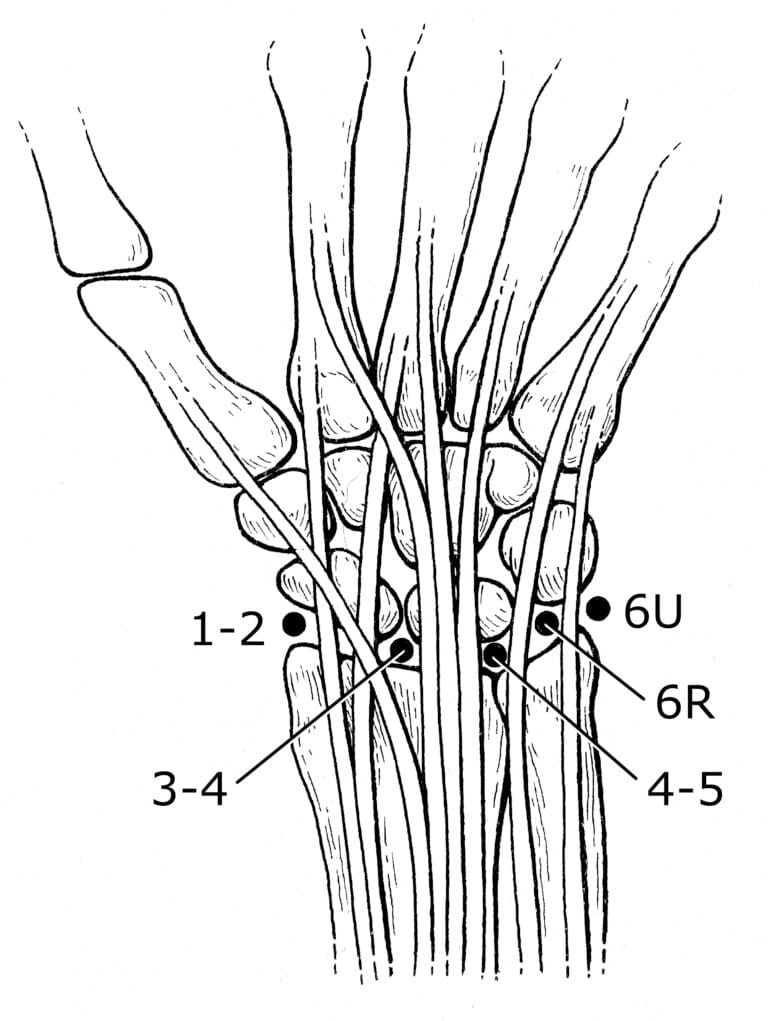
During the procedure, regional anesthesia is typically used to numb the hand and arm. A traction device may be applied to gently widen the wrist joint, making it easier to insert the arthroscope. The surgeon then creates small portals around the wrist to access the joint and may fill the area with saline to enhance visibility.

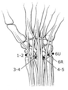
Surgeons place portals in specific locations on the wrist, depending on the area that needs to be viewed and treated. This set of portals, called the standard radiocarpal portals, will give access to the major joint(s) at the wrist. Other sets of portals provide access to other areas of the wrist.
Diagnostic Wrist Arthroscopy
Diagnostic wrist arthroscopy is a technique that uses an arthroscope to identify potential issues within the joint causing symptoms. It is often considered when:
- The source of wrist pain is not clearly identifiable through a physical exam, imaging tests such as X-rays, MRI scans, or other diagnostic tools.
- Persistent wrist pain continues for several months despite nonsurgical treatments.
If problems are found during the diagnostic procedure, they can often be treated arthroscopically at the same time, eliminating the need for additional surgery.
After the procedure, small incisions are closed with stitches, and a dressing is applied. In some cases, a splint may be used to support the wrist during the healing process.
Arthroscopic Surgical Treatments for Wrist Conditions
Arthroscopic surgery is a versatile technique used to address various wrist conditions, including:
- Chronic Wrist Pain: When other diagnostic tests fail to pinpoint the source of chronic wrist discomfort, exploratory arthroscopy can be used to find areas of inflammation, cartilage damage, or other abnormalities. Treatment can often be performed arthroscopically during the same procedure.
- Wrist Fractures: Arthroscopy helps remove small bone fragments from the joint and realign broken bones after a fracture. The stabilized bone may be fixed using pins, wires, screws, or plates.
- Ganglion Cysts: These fluid-filled lumps often form from a stalk between two wrist bones. Arthroscopy allows for the removal of the cyst and its stalk, effectively eliminating the cyst.
- Ligament and Triangular Fibrocartilage Complex (TFCC) Tears: The TFCC stabilizes and cushions the wrist, while ligaments connect bones to provide joint stability. Trauma, such as falling on an outstretched hand, can tear ligaments or the TFCC, resulting in pain or clicking. Arthroscopy can diagnose and repair these tears, restoring wrist function.
By incorporating minimally invasive techniques, arthroscopic wrist surgery enhances precision in diagnosing and treating various joint conditions, promoting quicker recovery and fewer complications.
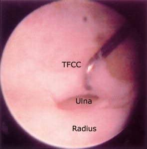
This is a view inside a wrist joint using an arthroscope. The TFCC ligament is torn, and the surgeon is using a hook to identify the tear. Afterward, the surgeon will use a shaver to clean the area to make a smooth surface at the tear. In some cases, a suture can then be passed to repair the tear. The head of the ulna is visualized through the tear. Next to it is the radius.
Post-Surgery Care
In the initial 2 to 3 days following wrist arthroscopy, it is crucial to keep the wrist elevated and ensure the bandage remains clean and dry. Applying ice can help reduce swelling.
To aid recovery, specific exercises may be recommended to maintain wrist mobility and restore strength. While post-operative pain is typically mild, analgesic medications can be used to manage discomfort. Your surgeon will provide a detailed plan for post-surgery pain relief tailored to your needs.
Potential Complications
Although complications from arthroscopic wrist surgery are rare, some risks exist, including:
- Infection
- Nerve damage
- Significant swelling
- Bleeding
- Scarring
- Tendon injuries
Your surgeon will discuss these potential complications with you before the procedure to ensure you are fully informed.
Expected Outcomes
The success of wrist arthroscopy largely depends on the condition diagnosed and treated during the procedure. However, most cases result in favorable outcomes, offering effective diagnosis and treatment for various wrist conditions


