Understanding Pediatric Ankle Fractures: Causes, Treatment, and Recovery

Pediatric ankle fractures, commonly referred to as broken ankles, are among the most frequent injuries in children. These fractures involve a break in one or more bones of the ankle joint: the tibia (shinbone), the fibula (outer ankle bone), or the talus (a small bone located at the back of the foot).

The severity of ankle fractures in children can vary widely. Minor fractures are often effectively treated with a simple walking boot, while complex fractures may necessitate surgical intervention. A pediatric orthopedic specialist will guide you through the available treatment options and provide ongoing follow-up care to ensure proper healing and monitor long-term recovery outcomes.

Anatomy of the Ankle Joint

The ankle joint is the point where the tibia (shinbone), fibula (outer ankle bone), and talus (a small bone in the foot) come together. These bones are held in place by ligaments, which function like sturdy ropes, providing stability to the joint by connecting bones and preventing excessive movement. The ankle contains several important ligaments that work together to maintain joint stability and facilitate smooth movement.
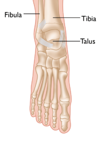
 

The normal skeletal anatomy of the foot and ankle.

Understanding Ankle Fractures in Children

In children and adolescents, ankle fractures typically affect the tibia or fibula, often involving the growth plates—specialized areas of cartilage near the ends of long bones. Growth plates play a critical role in bone development, gradually hardening into solid bone as the child matures. However, because these growth plates are the last parts of the bones to solidify, they are particularly prone to injury and fractures.

As adolescents approach the end of their growth phase, growth plates begin to close and harden, making them susceptible to transitional fractures during this maturing stage. Two common types of transitional ankle fractures are triplane fractures and Tillaux fractures, which require specific treatment approaches depending on their severity and location (refer to the Treatment section for more details)

 

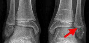
This X-ray of a child’s ankle taken from the front clearly shows the growth plates of the tibia and fibula (red arrows).

Causes of Pediatric Ankle Injuries

Ankle injuries in children often result from sudden twisting of the lower leg or foot, frequently occurring during sports or active play. High-energy activities, especially those involving lateral movements and jumping, significantly increase the risk of such injuries.

Sports like basketball, for instance, pose a higher risk because players often jump to defend, shoot, or rebound. If a child lands awkwardly or on another player’s foot, it can cause the ankle to twist or roll inward or outward, leading to potential fractures or sprains. Recognizing these risks can help in adopting preventative measures to minimize injury during physical activities.


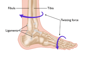
A twisting force to the lower leg or foot is a common cause of ankle fractures, as well as ligament injuries (sprains).

Symptoms of Pediatric Ankle Fractures

Differentiating between an ankle sprain and a fracture can be challenging, as both conditions initially cause pain and swelling. However, fractures often present additional signs and symptoms, such as:

  • Inability to stand or walk
  • Bruising
  • Tenderness to the touch
  • Visible deformity in the ankle area

Doctor Examination

Medical History and Physical Examination

A thorough evaluation begins with the doctor discussing your child’s medical history and the circumstances of the injury. This is followed by a careful physical examination of the ankle to assess:

  • Swelling and bruising
  • Tenderness at the injury site
  • Any deformity or misalignment of the ankle
  • Presence of tears or open wounds in the skin
  • Proper functioning of arteries and nerves to rule out further complications

Imaging Tests

When a fracture is suspected, imaging tests are crucial for an accurate diagnosis:

  • X-rays: The most commonly used tool for diagnosing fractures, X-rays provide clear images of the bone structure, helping determine whether a bone is broken.
  • Computed tomography (CT): A CT scan creates detailed cross-sectional images of the ankle bones, which are especially helpful for fractures that extend into the ankle joint or require surgical intervention.
  • Magnetic resonance imaging (MRI): In cases where a fracture is suspected but not visible on X-rays, an MRI may be ordered. MRI scans provide high-resolution images of both bones and soft tissues, such as ligaments, offering a comprehensive view of the injury.

These diagnostic steps ensure that your child’s condition is thoroughly evaluated, enabling precise treatment and a better recovery outcome.

Treatment of Pediatric Ankle Fractures

The treatment approach for ankle fractures in children depends on several key factors, including:

  • The specific location of the fracture
  • The extent of damage to the growth plate
  • The position of the foot at the time of injury
  • The direction and magnitude of the force causing the injury
  • The degree of bone fragment displacement (shifting)

Fractures involving growth plates require careful management due to potential long-term complications, such as improper ankle alignment or differences in leg length as the child grows.

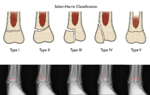
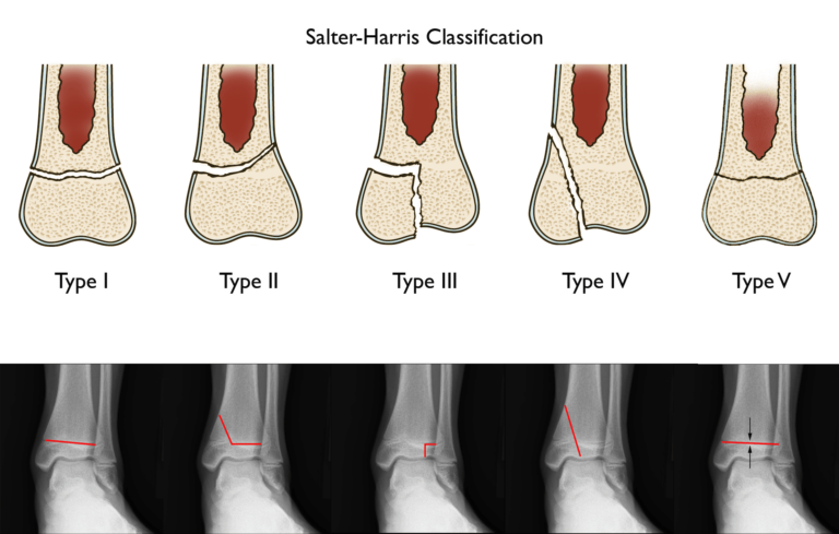
A widely recognized method for categorizing growth plate fractures is the Salter-Harris classification system. This system evaluates the severity of damage to the growth plate and assesses the risk of future growth disturbances. In this context, the Salter-Harris classification is used to describe various types of fractures and their corresponding treatment strategies, ensuring appropriate care tailored to the injury’s characteristics.


(Above) Standard illustrations of growth plate fracture types. (Below) The red lines in this X-ray of the ankle show the different types of growth plate fractures as they appear in the ankle joint.

Types of Pediatric Ankle Fractures and Treatment Approaches

Type I and Type II Fractures

Type I fractures involve a break through the growth plate, completely separating the bone end from the bone shaft and disrupting the growth plate entirely. Type II fractures extend through part of the growth plate and into the bone shaft. Both types are generally treated without surgery and have favorable outcomes with proper immobilization.

Type III and Type IV Fractures

Type III and Type IV fractures are more complex and have a higher risk of causing growth disturbances, potentially leading to an uneven or crooked ankle. Achieving precise bone alignment is crucial in these cases. Surgical intervention is often required to realign and stabilize the fracture for optimal healing and growth.

Type V Fractures

Type V fractures are typically identified after healing when complications such as growth disturbances become evident. These fractures are rare and can result in significant long-term issues if not properly managed.

Non-Displaced Fractures

When a fracture is well-aligned, stabilization with a walking boot or cast is sufficient to ensure proper healing. Most non-displaced fractures heal within 4 to 6 weeks, with the child’s doctor providing guidance on when weight-bearing can safely resume.

Displaced Fractures

Displaced fractures, where bone fragments are misaligned, require more intensive treatment. The first step is a closed reduction, a procedure to realign the bones without surgery, often performed in the emergency or operating room under sedation. If alignment is successfully restored, a cast is applied to maintain the proper position.

For fractures that cannot be properly aligned through closed reduction—commonly Type III and Type IV fractures involving the ankle joint—open reduction surgery is necessary. This surgical approach allows direct visualization of the bone fragments for precise alignment, especially when soft tissues like muscles interfere.

To maintain alignment, surgeons may use metal plates or screws. These internal fixation devices add stability, reducing the need for external immobilization. For instance, a walking boot may suffice for mobility if screws are used, whereas a cast would be required without them.

Distal Fibular Fractures

Isolated fractures of the distal fibula, often classified as Salter-Harris Type I or II, typically result from low-energy trauma, such as a fall. These fractures usually heal well with treatment using a walking boot or short-leg cast.

Triplane Fractures

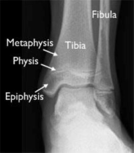
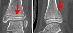
Triplane fractures are complex injuries seen in older adolescents whose growth plates are beginning to close. These fractures, categorized as Salter-Harris Type IV, span three planes: the metaphysis (widened part of the bone shaft), the physis (growth plate), and the epiphysis (bone end). Due to their complexity, triplane fractures require detailed imaging and may often need surgical alignment to ensure proper healing.


The distal tibia of a growing child comprises three sections: the physis, epiphysis, and metaphysis.

Treatment of Triplane Fractures

The treatment approach for triplane fractures largely depends on the degree of displacement between the bone fragments. For minimally displaced or non-displaced triplane fractures, immobilization with a long-leg cast is typically sufficient to ensure proper healing.

However, triplane fractures are often displaced, requiring more advanced intervention. In such cases, open reduction and internal fixation (ORIF) using screws is the preferred method. This surgical technique allows for precise realignment of the bone fragments and helps maintain the reduction throughout the healing process, improving the likelihood of a successful recovery.


This CT scan of the ankle taken from the front (left) and from the side (right) shows a triplane fracture. Note the fracture plane is different in each view. The fracture also runs across the growth plate, resulting in three fracture planes (triplane).

Tillaux Fractures: Causes, Characteristics, and Treatment

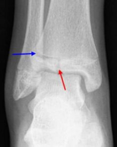
Tillaux fractures, named after the French surgeon Paul Tillaux, are a specific type of Salter-Harris Type III fracture that occurs in the front and outer portion of the lower tibia. These fractures extend through the growth plate and into the ankle joint, representing approximately 3% to 5% of pediatric ankle fractures.

Tillaux fractures typically arise in late adolescence, a period when the growth plate begins to close. Due to this transitional phase, the fracture fragments are often loose and unstable, requiring specialized treatment.

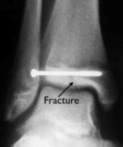
The most effective treatment for Tillaux fractures is open reduction and screw fixation, a surgical approach that ensures precise alignment and stability of the bone fragments. This method promotes proper healing and minimizes the risk of long-term complications, such as joint instability or growth disturbances.


In this Tillaux fracture, the blue arrow shows a widening of the growth plate, and the red arrow shows the fracture extending through the epiphysis and into the joint.


During surgery to treat this Tillaux fracture, the bone fragments have been set into alignment and held together with a screw fixation.

Medial Malleolus Fractures: Overview and Treatment

The medial malleolus is the prominent bony bump located on the inside of the ankle. In children, a medial malleolus fracture can occur when the ankle is twisted, often resulting in a small and unstable bone fragment.

These fractures are more common in younger children, where the risk of complications from a growth disturbance is higher due to the ongoing development of the growth plate.

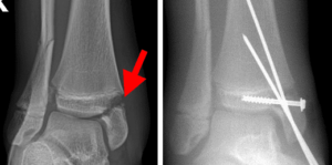
For non-displaced or minimally displaced fractures, immobilization with a long-leg cast is generally effective. However, because the fracture fragment is typically small and unstable, many cases require screw and/or pin fixation to ensure proper alignment and stability during the healing process.


This X-ray shows a fractured and displaced medial malleolus. The bone fragment was reduced to its normal position and stabilized with a screw and two pins. Screw fixation alone is typically adequate.

Recovery and Complications of Pediatric Ankle Fractures

Recovery Timeline and Care

Most ankle fractures in children heal within 4 to 6 weeks, with pain typically subsiding significantly as healing progresses. The use of a splint, cast, or surgical stabilization helps reduce discomfort, and most children experience minimal pain within two weeks of the injury. To manage any remaining pain, doctors may prescribe appropriate pain medications.

For mild fractures, walking may be permitted early on, but in most cases, weight-bearing is restricted until the fracture fully heals. Your child’s doctor will provide personalized guidance on when weight-bearing activities can resume.

Although the fracture itself often heals in 4 to 6 weeks, complete recovery takes longer. Rehabilitation may involve home exercises or physical therapy to restore joint flexibility and leg muscle strength. Returning to normal activities is typically recommended only after full strength and mobility are regained.

Potential Complications

Pediatric ankle fractures, particularly those involving the growth plate, carry a risk of growth disturbances. These complications can result in a crooked ankle or one leg growing shorter than the other. Because of this, careful monitoring by a doctor is essential to ensure proper healing and growth.

Follow-up visits should continue for at least one year after the injury to evaluate the fracture’s impact on growth. In cases of more severe or complicated fractures, monitoring may need to extend until the child reaches skeletal maturity to prevent or address long-term issues.


In this X-ray, the growth has stopped on the patient’s left fibula after the fracture healed (arrow). Compare this to the fibular growth plate on the uninjured side of the other ankle. This has resulted in some shortening of that bone and pain in the involved ankle.

Conclusion

Ankle fractures are a frequent injury among children and adolescents. When properly treated and well-aligned during healing, these fractures typically resolve without leading to significant long-term complications, such as arthritis or chronic pain. Ensuring timely medical care and monitoring, particularly for fractures involving the growth plate, helps prevent future issues and supports a full recovery.

Scroll to Top