Understanding Hip Fractures: Causes, Risks, and Treatment Approaches

A hip fracture refers to a break in the upper segment of the femur, commonly known as the thighbone. These fractures are most frequently seen in older adults with weakened bones due to osteoporosis. However, in younger individuals, hip fractures typically result from high-impact incidents, such as falling from a significant height or being involved in a vehicular accident.

Annually, over 300,000 individuals in the United States experience hip fractures, with the majority being adults aged 65 and older. These injuries predominantly occur during falls at home or within the community.

Hip fractures are often associated with significant pain, making timely surgical intervention crucial. Early treatment not only addresses the fracture but also enables patients to mobilize quickly, reducing the risk of complications such as bed sores, blood clots, and pneumonia. Prolonged immobility in elderly patients can lead to further issues, including cognitive disorientation, which complicates rehabilitation and recovery efforts.


Most hip fractures sustained by older people result from falls.

Anatomy of the Hip Joint

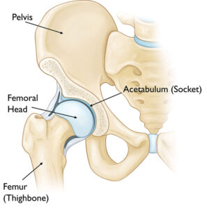
The hip joint is a classic example of a ball-and-socket structure.

  • The Ball: This is the femoral head, which forms the uppermost portion of the thighbone (femur).
  • The Socket: Known as the acetabulum, this part of the pelvis has a rounded shape designed to securely encase the femoral head. Together, this structure allows for a wide range of motion while maintaining joint stability.


Normal hip anatomy

Detailed Description of Hip Fractures

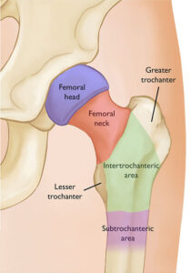
A hip fracture involves damage to one of the four key regions of the upper femur:

  1. Femoral Neck: Located just beneath the femoral head (the ball), this area connects the head to the shaft of the femur.
  2. Intertrochanteric Region: This is the area between the femoral neck and the shaft, defined by two prominent bony structures — the greater and lesser trochanters.
  3. Subtrochanteric Region: The part of the femoral shaft just below the greater and lesser trochanters.
  4. Femoral Head: The ball-shaped top of the femur that fits snugly within the acetabulum.

Among these, intertrochanteric fractures and femoral neck fractures are the most frequently occurring types of hip fractures. On the other hand, femoral head fractures are exceedingly rare, often resulting from high-impact injuries like car accidents or severe falls.

This comprehensive understanding of hip anatomy and the common fracture types can aid in identifying the nature of the injury and tailoring appropriate treatment plans.


The areas of the femur (thighbone). Most hip fractures occur in the femoral neck or intertrochanteric area.

Causes of Hip Fractures

In elderly individuals with weakened or osteoporotic bones, most hip fractures result from low-energy falls. Even minor incidents, such as twisting awkwardly or tripping, can lead to fractures.

In some instances, the bone’s fragility may cause it to fracture spontaneously during routine activities like walking or standing. This phenomenon is often described as “the break occurs before the fall,” with such fractures typically occurring in the femoral neck.

Stress fractures, caused by repeated impact, are another potential cause and are most commonly observed in the femoral neck. These injuries are prevalent among long-distance runners and military recruits undergoing intense physical training. Stress fractures in the subtrochanteric region may also occur due to prolonged use of specific osteoporosis medications.

On the other hand, femoral head fractures are rare and generally result from high-impact trauma or as part of a hip fracture dislocation.

Symptoms of Hip Fractures

Hip fractures are usually marked by sudden, intense pain localized to the groin or upper thigh. In most cases, individuals cannot stand, bear weight, or move their upper leg or knee. Movement of the ankle and toes is generally unaffected unless there are additional injuries to the lower leg.

In rare instances, some fractures may allow partial weight-bearing, though it will still cause severe pain. Nondisplaced fractures of the femoral neck, in particular, may not immediately present with intense pain, and some individuals might still manage limited movement.

Doctor Examination

Physical Assessment
Most patients with suspected hip fractures are brought to the emergency room for evaluation. Here, an emergency physician or orthopedic surgeon will assess the injury, take a detailed history, and check for other potential injuries. A thorough examination of sensation, movement, and circulation in the lower leg is also performed.

Common findings include:

  • The injured leg appearing shorter than the other.
  • Twisting or rotation of the leg, either internally or externally.
  • Bruising on the outer hip or thigh at the point of impact.
  • Limited and painful movement of the leg.

In cases of nondisplaced fractures of the femoral neck, the pain may be less severe, prompting patients to visit a doctor’s office instead of the emergency room. These fractures may still allow limited weight-bearing, albeit with discomfort.

Imaging Studies for Diagnosis

Imaging studies play a vital role in diagnosing and understanding the extent of hip fractures.

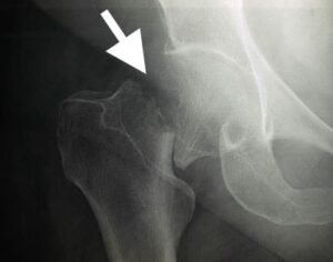
  • X-rays: Provide clear images of dense structures like bones and are typically sufficient to identify most hip fractures.


Most hip fractures are visible on an x-ray.


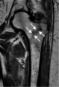
  • Magnetic Resonance Imaging (MRI): Offers detailed views of both soft tissues and bone, making it especially useful for detecting small or incomplete fractures that may not appear on X-rays.


This MRI scan shows a hip fracture that was not visible on an x-ray.


  • Computerized Tomography (CT): Provides cross-sectional images of the hip, offering additional insights into complex fractures that require detailed evaluation.

Accurate imaging ensures proper diagnosis and guides the treatment plan, improving outcomes for individuals with hip fractures.


Scroll to Top