Thoracic Disc Herniation

Introduction

The increased use of magnetic resonance imaging (MRI) has led to the frequent detection of thoracic disc herniations, with studies estimating that up to 15% of the population may have this condition. However, in many cases, these herniations are incidental findings, appearing on MRI scans conducted for unrelated issues.

Most individuals with thoracic disc herniation remain asymptomatic and do not experience significant problems. When symptoms do occur, the primary concern is whether the herniated disc is compressing the spinal cord. Despite common references to a “slipped disc,” the disc does not actually shift out of place; instead, herniation refers to the protrusion of the central disc material beyond its normal boundary. Thoracic disc herniation is most commonly observed in individuals aged 40 to 60 years.

This guide provides insights into:

• The underlying causes and progression of thoracic disc herniation
• How physicians diagnose the condition
• Available treatment options for managing symptoms

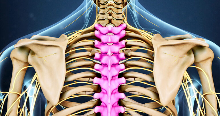
Anatomy of the Thoracic Spine

What Structures Are Involved?

The human spine consists of 24 vertebrae, stacked in a column to form the spinal column. The thoracic spine is composed of 12 vertebrae (T1-T12), extending from the base of the neck to the lower ribcage. The T12 vertebra serves as a transitional point, connecting the thoracic spine to the lumbar spine (L1).

• The upper thoracic spine (T1-T6) is relatively immobile due to its attachment to the ribcage, making disc herniations in this region uncommon.
• The lower thoracic spine (T8-T12) is more flexible and susceptible to mechanical stress, accounting for nearly 75% of thoracic disc herniations, particularly at T11-T12.

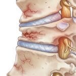
The intervertebral discs between the vertebrae provide shock absorption and stability. Each disc consists of:

• Nucleus pulposus – A gel-like central core responsible for cushioning forces.
• Annulus fibrosus – A strong outer layer of fibrous rings, securing the nucleus in place.

A herniation occurs when the nucleus pulposus protrudes through the annulus fibrosus, leading to compression of the spinal cord or nerve roots.

The Critical Zone: Why Some Herniations Are More Dangerous

The spinal canal houses the spinal cord, which extends through the thoracic spine. Unlike the cervical and lumbar regions, the thoracic spinal canal is narrower, making any space-occupying lesion, such as a herniated disc, a potential risk for spinal cord compression.

A particularly vulnerable region within the thoracic spine is the “critical zone” (T4-T9). This section is supplied by only one blood vessel—the anterior spinal artery. Compression in this area can compromise blood flow, leading to spinal cord ischemia and potential paralysis if untreated.

Causes of Thoracic Disc Herniation

Why Does This Condition Develop?

Thoracic disc herniation primarily results from degenerative changes in the spine. Over time, the intervertebral discs experience wear and tear, leading to:

• Disc dehydration and loss of elasticity, making them more prone to herniation.
• Small tears in the annulus fibrosus, allowing disc material to protrude.
• Progressive degeneration, particularly at T11-T12, due to mechanical forces from bending and twisting.

Other Contributing Factors

In some cases, a herniation may occur suddenly due to acute trauma. These events include:

• High-impact injuries – Such as falls or motor vehicle accidents.
• Repetitive strain – Chronic overuse from activities involving twisting, lifting, or poor posture.
• Pre-existing spinal conditions – Individuals with Scheuermann’s disease have a higher likelihood of multiple thoracic disc herniations.

When a herniation occurs in the critical zone (T4-T9), there is an increased risk of spinal cord injury due to direct compression or reduced blood flow. This can result in significant neurological deficits, including weakness, loss of sensation, and, in severe cases, paralysis.

Symptoms of Thoracic Disc Herniation

What Are the Signs and Symptoms?

Symptoms of thoracic disc herniation vary based on:

• The location and size of the herniation.
• Whether the disc is compressing the spinal cord or nerve roots.

Common symptoms include:

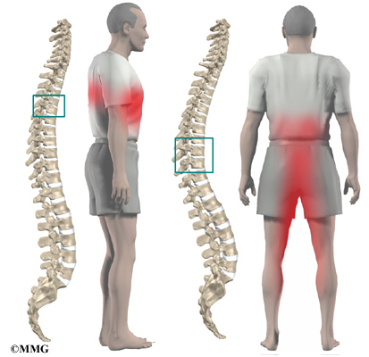
• Mid-back pain – The most frequent symptom, often worsened by movement, deep breathing, or prolonged sitting.
• Radiating pain – Discomfort may extend to the chest, ribs, or abdomen, sometimes mimicking heart, lung, or gastrointestinal conditions.
• Numbness and tingling – Sensory disturbances can occur along the rib cage or lower extremities.
• Muscle weakness – In cases of spinal cord compression (myelopathy), patients may experience leg weakness, difficulty walking, or even bowel and bladder dysfunction.

Referred Pain: How Thoracic Disc Herniation Mimics Other Conditions

Depending on the level of disc herniation, symptoms may radiate to different parts of the body, making diagnosis challenging:

• Upper thoracic herniations (T1-T5) – May cause pain in the arms, chest, or mimic cardiac symptoms.
• Mid-thoracic herniations (T6-T9) – Can lead to abdominal pain, sometimes mistaken for gastrointestinal issues.
• Lower thoracic herniations (T10-T12) – May result in groin or lower limb pain, resembling kidney disorders.

If the spinal cord is compressed, patients may experience progressive neurological decline, requiring urgent medical evaluation.

Diagnosis and Treatment of Thoracic Disc Herniation

Diagnosis

How Is Thoracic Disc Herniation Diagnosed?

The diagnostic process begins with a comprehensive medical history and physical examination. Your doctor will ask about the nature and severity of your symptoms, including pain distribution, numbness, weakness, and any issues with bowel or bladder function. Additionally, they will assess which movements or activities exacerbate or relieve symptoms.

A physical examination follows, evaluating range of motion, muscle strength, reflexes, and sensory function to identify any neurological deficits associated with the herniation.

Imaging Studies

• X-rays: While X-rays primarily visualize bone structures, they can sometimes detect calcified thoracic discs, which may indicate herniation. The cause of disc calcification is not fully understood but may be linked to prior injury.

• Magnetic Resonance Imaging (MRI): MRI is the gold standard for diagnosing thoracic disc herniation. This imaging modality provides high-resolution images of the spinal soft tissues, allowing for clear visualization of herniated discs and their impact on the spinal cord and nerve roots.

• Myelography and Computed Tomography (CT): Before the advent of MRI, myelography—an X-ray study involving the injection of contrast dye into the spinal canal—was commonly used. Today, CT myelography is sometimes utilized in surgical planning, offering detailed visualization of spinal cord compression and bony structures.

Most diagnoses rely on MRI, with CT scans and myelography reserved for complex cases or preoperative assessments.

Treatment Options for Thoracic Disc Herniation

Non-Surgical Treatment

In many cases, thoracic disc herniation can be managed without surgery, provided that there is no severe spinal cord compression or rapidly worsening neurological symptoms. The primary goal is to control pain, reduce inflammation, and restore mobility.

Conservative Management Strategies

• Activity Modification and Immobilization:
Temporary restriction of movement may help alleviate symptoms. A short period of bed rest (one to two days) can relieve pressure on the affected disc, but prolonged inactivity is discouraged to prevent muscle deconditioning. A thoracic brace may also provide temporary support.

• Medications:

  • Nonsteroidal anti-inflammatory drugs (NSAIDs) such as ibuprofen or aspirin help reduce pain and inflammation.
  • Muscle relaxants may be prescribed if muscle spasms contribute to discomfort.
  • Oral corticosteroids are sometimes used in a tapering dose to manage severe pain radiating into the limbs.

• Physical Therapy:
A structured rehabilitation program supervised by a physical therapist is crucial in maintaining spinal flexibility, improving posture, and preventing recurrence. Therapy typically involves:

  • Postural training to reduce spinal stress
  • Strengthening exercises for core and back muscles
  • Stretching routines to enhance mobility
  • Pain management techniques, including manual therapy and modalities such as heat or electrical stimulation

Most patients with thoracic disc herniation improve within six weeks of nonoperative care. If symptoms persist or worsen, surgical intervention may be considered.

Surgical Treatment

When Is Surgery Necessary?

Surgical intervention is recommended when:
• Non-surgical treatments fail to provide relief after six weeks
• Spinal cord compression results in neurological deficits, such as muscle weakness, numbness, or bowel and bladder dysfunction
• The condition rapidly progresses, posing a risk of permanent nerve damage

Several surgical approaches are available depending on the location and severity of the herniation.

Common Surgical Procedures

1. Costotransversectomy and Discectomy

This posterior approach involves the removal of a portion of the rib (costo-) and transverse process (-ectomy) to create an opening for disc removal. A small section of the damaged disc that is compressing the spinal cord is excised using specialized instruments. Care is taken to preserve spinal stability while decompressing the nerve structures.

2. Transthoracic Decompression

In transthoracic decompression, the surgeon accesses the herniated disc through the chest cavity. This method provides a clear, direct view of the disc, allowing for precise removal of the herniated material.

Procedure:

  • The patient is positioned on their side.
  • A small incision is made between the ribs.
  • Instruments are inserted to remove the herniated disc and relieve pressure on the spinal cord.

This anterior approach is beneficial for larger or centrally located herniations.

3. Video-Assisted Thoracoscopic Surgery (VATS)

A minimally invasive alternative to traditional open surgery, VATS utilizes a tiny camera (thoracoscope) inserted through a small incision in the thorax. The surgeon views the spinal structures on a monitor while removing the herniated disc.

Advantages of VATS include:

  • Smaller incisions
  • Reduced postoperative pain
  • Faster recovery
  • Less scarring around nerves and joints

4. Spinal Fusion (Arthrodesis)

If a significant portion of the disc or vertebrae is removed, spinal instability may occur, necessitating fusion.

Procedure:

  • Bone grafts are placed between vertebrae to encourage fusion.
  • Metal screws, rods, or plates may be used for additional stability.

Fusion is not always required but may be recommended if a large amount of disc or bone material is removed.

Conclusion

Thoracic disc herniation is often asymptomatic but may cause significant pain, neurological symptoms, or spinal cord compression in some cases. Non-surgical treatment is the first line of management and is effective in most patients. However, if symptoms worsen or fail to improve, surgical options, including costotransversectomy, transthoracic decompression, VATS, and spinal fusion, may be necessary to relieve pressure on the spinal cord and nerves.

Early diagnosis and a comprehensive treatment approach can significantly improve outcomes and prevent long-term complications.

Rehabilitation and Recovery After Thoracic Disc Herniation Treatment

Nonsurgical Rehabilitation

For patients who do not require surgery, a structured rehabilitation program is often recommended to manage symptoms and restore spinal function. A physical therapist typically guides the rehabilitation process, with sessions scheduled a few times per week for four to six weeks.

Early-Stage Rehabilitation Goals:

• Pain Management: Strategies such as postural training, heat or ice application, and ergonomic modifications help alleviate discomfort.
• Safe Movement Education: Patients learn proper body mechanics to reduce strain on the spine during daily activities.
• Gentle Mobility Exercises: Movements that promote spinal flexibility while avoiding excessive strain are introduced.

Progressive Rehabilitation:

As symptoms improve, the focus shifts to gradual strengthening through:
• Aerobic exercises (e.g., walking, swimming) to enhance endurance and overall well-being.
• Core and spinal stability exercises to improve back support and posture.

Most patients experience significant improvement within this timeframe. However, continued adherence to prescribed exercises is essential for long-term spinal health.

Rehabilitation After Surgery

Postoperative rehabilitation varies depending on the type of surgical procedure performed and individual patient factors.

Immediate Post-Surgical Care:

Some patients are discharged shortly after surgery, while others may require a hospital stay for a few days. During hospitalization, early mobilization is encouraged with the assistance of a physical therapist, who helps patients regain mobility while minimizing strain on the spine.

Early Recovery Guidelines:

• Use of a back brace or support belt may be required, depending on the surgeon’s recommendations.
• Avoid excessive activity during the initial healing phase to prevent complications.

Outpatient Physical Therapy:

Following discharge, many patients continue rehabilitation for one to three months under professional supervision.

Early-Stage Therapy Focus:
• Pain relief techniques, such as heat therapy, electrical stimulation, ultrasound, and massage, may be used to reduce discomfort and muscle spasms.
• Gentle mobility exercises for the hips, shoulders, and spine help restore movement.

Progression to Strengthening and Endurance Training:
As recovery advances, patients engage in:
• Strengthening exercises targeting the back and core muscles to support spinal stability.
• Water therapy (hydrotherapy) to facilitate movement with minimal stress on the spine.
• Postural correction training to improve body alignment and prevent future injury.

Patients gradually progress to more advanced exercises as their strength, endurance, and function improve.

Long-Term Recovery and Maintenance

The goal of rehabilitation is to return patients to their prior level of activity safely. While many individuals resume normal activities, some may need to modify their routines to minimize the risk of recurrent injury.

As formal therapy sessions conclude, patients are expected to continue a structured home exercise program to maintain flexibility, strength, and spinal health. Regular physical activity and adherence to good posture practices play a critical role in long-term success and symptom prevention.

By following a well-structured rehabilitation plan, patients can achieve optimal recovery and reduce the likelihood of future spinal complications.


Scroll to Top