Osteogenesis Imperfecta in Children: Understanding the Genetic Bone Disorder
Introduction
Osteogenesis Imperfecta (OI), commonly known as “brittle bone disease,” is a rare genetic disorder that primarily affects the bones, making them unusually fragile and prone to fractures. This condition stems from a defect in the production or quality of collagen, particularly Type I collagen, which is a crucial component of bone structure and strength. Although it affects individuals across all age groups, the manifestations in children are particularly significant due to their growth and developmental needs.
In this detailed article, we will explore the nature of Osteogenesis Imperfecta in children, delve into its genetic and molecular foundations, discuss its clinical presentations, diagnostic approaches, and available management options. We will also examine its impact on quality of life, rehabilitation strategies, and the latest advancements in research and treatment.
What is Osteogenesis Imperfecta?
Osteogenesis Imperfecta is a heterogeneous group of inherited disorders characterized by varying degrees of bone fragility and deformity. The hallmark of OI is an increased susceptibility to fractures with minimal or no trauma. However, the disorder encompasses a broader clinical spectrum, including joint laxity, hearing loss, dental anomalies, and scoliosis.
The condition is genetically diverse, and its severity can range from mild forms with few fractures to perinatal lethal types where infants may not survive beyond birth due to multiple fractures and underdeveloped lungs.
The Genetic Basis of OI in Children
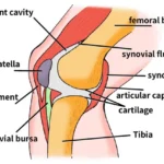
Collagen, particularly Type I collagen, forms the backbone of the bone matrix. It acts as a scaffold, around which minerals like calcium phosphate are deposited, contributing to bone rigidity and strength. In children with OI, mutations in the genes responsible for producing collagen result in abnormal collagen synthesis. This leads to structurally weak bones that fracture easily.
The majority of OI cases are associated with mutations in the COL1A1 and COL1A2 genes, which encode the alpha chains of Type I collagen. These mutations can result in either reduced production of collagen (quantitative defect) or production of structurally abnormal collagen (qualitative defect).
OI is primarily inherited in an autosomal dominant pattern, though autosomal recessive and sporadic mutations have also been documented. This means that a child has a 50% chance of inheriting the condition from an affected parent in dominant inheritance.
Classification of Osteogenesis Imperfecta
Over the years, the classification of OI has evolved. The most commonly used system is the Sillence Classification, which identifies four primary types based on severity and clinical presentation. More recently, expanded classifications have identified additional types (Types V–XVIII), incorporating rare genetic variants.
Sillence Types Overview:
– Type I (Mild): Most common and least severe. Normal stature, blue sclerae, and a tendency for fractures.
– Type II (Perinatal Lethal): Most severe. Multiple fractures at birth, respiratory failure due to underdeveloped lungs.
– Type III (Severe): Progressive deformity, short stature, frequent fractures from early infancy.
– Type IV (Moderate): Intermediate severity. Normal sclerae, moderate bone deformity.
Clinical Features and Symptoms in Children
Children with OI often present with frequent fractures, sometimes from normal handling or minimal trauma. Bone deformities such as bowed limbs, scoliosis, or vertebral compression fractures may be observed.
Short stature and growth retardation are common, particularly in the more severe forms. Delays in gross motor development are also frequently noted due to fracture risk and limited mobility.
One of the hallmark signs, especially in Type I OI, is the presence of bluish discoloration of the sclerae (whites of the eyes), resulting from the thinness of the collagen matrix in the eyes.
A significant number of children with OI also suffer from dental problems, including discolored, brittle, or misshaped teeth, known as dentinogenesis imperfecta.
Progressive hearing impairment, often beginning in adolescence or early adulthood, is another complication. It may be due to abnormalities in the middle and inner ear bones.
Children may experience loose joints (hyperlaxity) and reduced muscle tone, which further complicate mobility and physical activities.
Diagnosis of Osteogenesis Imperfecta in Children
Diagnosis of OI typically begins with a thorough clinical history and physical examination. Key indicators include:
– History of frequent fractures
– Blue sclerae
– Dental abnormalities
– Family history of similar symptoms
Pediatricians often suspect OI early, especially in cases where multiple fractures occur without adequate trauma.
X-rays reveal characteristic features such as:
– Multiple healed fractures at various stages
– Bone deformities like bowing or shortening
– Thin cortical bones and reduced bone density
– Wormian bones in the skull
In severe types, prenatal ultrasound may detect fractures or shortened limbs before birth.
Molecular genetic testing can identify mutations in the COL1A1, COL1A2, or other collagen-related genes. This not only confirms the diagnosis but also helps determine the type and inheritance pattern.
Dual-energy X-ray absorptiometry (DEXA) is used to assess bone mineral density, which is often markedly low in children with OI.
Treatment and Management
There is currently no cure for Osteogenesis Imperfecta, but multidisciplinary treatment strategies can significantly improve quality of life and reduce complications.
Pharmacological Treatment:
– Bisphosphonates: These drugs, such as pamidronate and zoledronic acid, are the cornerstone of OI treatment. They increase bone mineral density, reduce fracture rates, and improve mobility in children.
– Growth Hormone Therapy: In selected cases, growth hormone therapy may aid in increasing stature and bone mass, although its use remains controversial and limited to specific cases.
Surgical Interventions:
– Intramedullary Rodding: A common orthopedic procedure in children with OI is telescopic rodding, where flexible rods are inserted into long bones (femur, tibia) to prevent or correct deformities and support bone growth.
– Other procedures may include spinal fusion surgery for scoliosis and fracture fixation or correction of deformities.
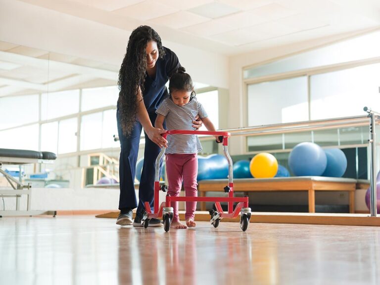
Physical Therapy and Rehabilitation:
A key component of care, physical therapy helps improve:
– Muscle strength and tone
– Joint stability
– Mobility and gait
Customized rehabilitation plans focus on developing independence in movement while minimizing fracture risk.
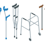
Orthotic Devices and Mobility Aids:
Children may benefit from braces, walkers, wheelchairs, or customized footwear to support movement and reduce stress on fragile bones.
Nutritional Support and Lifestyle Management
Proper nutrition plays a vital role in managing OI:
– Adequate calcium and Vitamin D intake
– Balanced diet to support muscle and bone growth
– Avoiding sedentary lifestyles, while promoting safe, low-impact physical activity like swimming
Psychosocial and Educational Considerations
Mental Health Support:
Children with OI may face psychological challenges such as:
– Fear of injury
– Social isolation
– Depression or anxiety
Counseling and emotional support from caregivers and professionals is essential for coping and confidence-building.
School Participation and Accessibility:
Children with OI should be encouraged to attend mainstream schools, with accommodations such as:
– Mobility-friendly environments
– Assistance during physical education
– Extra care with seating arrangements and classroom transitions
Complications and Long-term Outcomes
Orthopedic Complications:
– Recurrent fractures and deformities
– Spinal curvature and vertebral collapse
– Premature arthritis in weight-bearing joints
Hearing Impairment and Dental Problems:
Both can progress over time and require audiology assessments and dentistry interventions.
Respiratory and Cardiovascular Complications:
Severe types of OI may involve restrictive lung disease, especially in cases with rib deformities or spinal scoliosis.
Prognosis and Quality of Life
The long-term prognosis for children with OI largely depends on:
– Type and severity of OI
– Timely interventions and therapy
– Supportive home and school environments
With modern treatments and assistive technologies, many children with mild to moderate OI lead independent, fulfilling lives.
Research and Future Directions
Gene Therapy:
Promising advancements in gene editing technologies like CRISPR-Cas9 aim to correct collagen gene mutations at the molecular level. Although still in early research phases, these approaches hold future potential for curative therapy.
Stem Cell Therapy:
Researchers are exploring mesenchymal stem cell transplantation, which may help improve collagen production and bone quality.
Targeted Drug Therapy:
Future treatments may focus on modulating bone resorption or enhancing collagen stability using newer pharmaceuticals.
Frequently Asked Questions (FAQs)
- Is Osteogenesis Imperfecta curable?
No, there is currently no cure, but treatment can greatly improve quality of life.• Can children with OI lead normal lives?
Yes, with proper treatment and support, many children can lead productive and independent lives.• Does OI worsen with age?
Not necessarily. Some children experience fewer fractures after puberty, but complications can persist.• Can OI be prevented?
As a genetic condition, OI cannot be prevented, but genetic counseling can help families understand risks.• Is physical activity safe for children with OI?
Yes, with precautions. Activities like swimming and hydrotherapy are often recommended.
Conclusion
Osteogenesis Imperfecta in children presents unique challenges but is manageable through a comprehensive, multidisciplinary approach. Early diagnosis, appropriate therapies, nutritional support, and psychological care play critical roles in improving the child’s overall health and development. Continued research brings hope for more targeted, effective treatments in the