Introduction
Adult forearm fractures are common injuries that can occur due to various reasons such as falls, direct blows, or motor vehicle accidents. The forearm consists of two bones, the radius and the ulna, which can break in several ways. Proper diagnosis, treatment, and rehabilitation are crucial for optimal recovery and function.
Anatomy of the Forearm
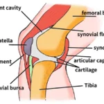
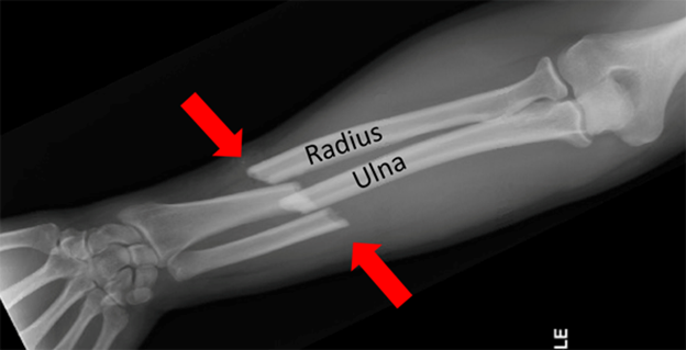
The forearm is made up of the radius and ulna, which run parallel to each other. These bones support the muscles that control the wrist and elbow, making them vital for hand and arm movements. The proximal ends of these bones form joints with the humerus at the elbow, while the distal ends form joints with the carpal bones at the wrist.
Types of Forearm Fractures
Forearm fractures can be classified based on the location and type of break:
- Distal Radius Fractures: These are the most common and occur near the wrist.
- Midshaft Fractures: These occur along the length of the forearm.
- Proximal Radius and Ulna Fractures: These occur near the elbow.
Fractures can also be categorized as:
- Simple Fractures: Where the bone breaks cleanly into two pieces.
- Comminuted Fractures: Where the bone is shattered into multiple pieces.
- Open (Compound) Fractures: Where the bone pierces the skin.
- Closed Fractures: Where the skin remains intact.
Causes and Risk Factors
Forearm fractures in adults often result from high-energy impacts, such as falls from a height, sports injuries, or car accidents. Risk factors include osteoporosis, advanced age, and conditions that affect bone strength and coordination.
Symptoms
Common symptoms of a forearm fracture include:
- Severe pain and tenderness
- Swelling and bruising
- Deformity or abnormal positioning of the forearm
- Inability to rotate the arm
- Numbness or tingling in the hand or fingers
Diagnosis
Diagnosis involves a physical examination and imaging tests. X-rays are the primary diagnostic tool, providing clear images of the bone structure. In some cases, a CT scan or MRI may be necessary to assess the extent of the injury and any associated soft tissue damage.
Treatment Options
Treatment depends on the type, location, and severity of the fracture, as well as the patient’s overall health and activity level.
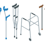
Non-Surgical Treatment:
- Casting and Splinting: For simple, non-displaced fractures, immobilization with a cast or splint is often sufficient.
- Bracing: Functional braces may be used to allow limited movement while maintaining alignment.
Surgical Treatment:
- Internal Fixation: Metal plates and screws are used to stabilize the bones.
- Intramedullary Nailing: A metal rod is inserted into the marrow canal of the bone.
- External Fixation: Pins are inserted into the bone and connected to an external frame to hold the bones in place.
Rehabilitation
Rehabilitation is essential for restoring function and strength. It involves physical therapy to improve range of motion, strength, and flexibility. Early movement of the fingers, wrist, and elbow is encouraged to prevent stiffness.
Complications
Potential complications include infection, non-union or malunion of the bones, nerve or blood vessel damage, and arthritis in the affected joints.
Advances in Treatment
Recent advancements in orthopedic surgery, such as minimally invasive techniques and improved fixation devices, have enhanced the outcomes for patients with forearm fractures. These innovations reduce recovery time and improve functional results.
Conclusion
Adult forearm fractures require timely and appropriate treatment to ensure optimal recovery. Understanding the types, causes, symptoms, and treatment options is crucial for effective management. Consultation with an orthopedic specialist is recommended for personalized care.