Overview:
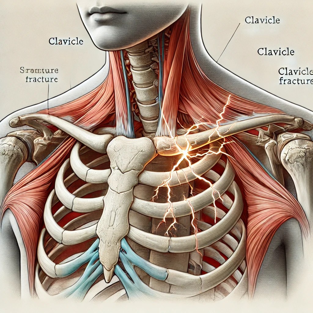
Clavicular fractures, commonly known as collarbone fractures, are frequent injuries, making up 2.6-5% of all fractures and 35-44% of shoulder girdle injuries. These fractures often occur due to direct impacts, falls on the shoulder, or sports-related incidents.
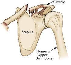
What is the Clavicle?
The clavicle, or collarbone, is a long bone that connects the shoulder blade to the breastbone (sternum). It plays a crucial role in shoulder movement and stability.
Causes of Clavicular Fractures:
- Direct Blows: A direct hit to the shoulder, often seen in contact sports like football or hockey.
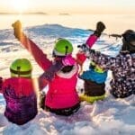
- Falls: Falling onto the shoulder or an outstretched arm can result in a fracture.
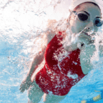
- Sports Injuries: High-impact sports activities increase the risk of breaking the clavicle.
Symptoms of a Clavicular Fracture:
- Pain: Sharp pain at the site of the fracture.
- Swelling and Bruising: Visible swelling and bruising around the collarbone.
- Deformity: A noticeable bump or deformity over the break.
- Limited Movement: Difficulty or pain when moving the arm and shoulder.
Diagnosing Clavicular Fractures:
- Physical Examination: The doctor will check for pain, swelling, and deformity.
- Imaging:
- X-rays: The primary tool to see the fracture and determine its type.
- CT Scans: Used for more detailed images, especially in complex cases.
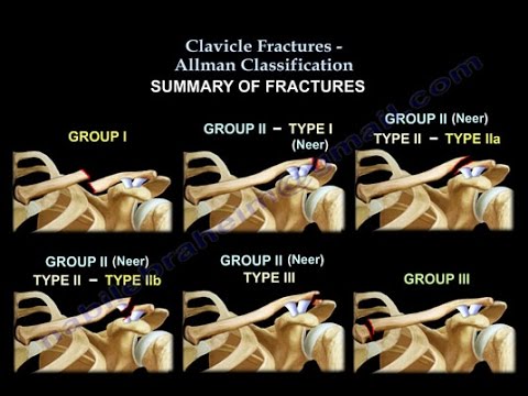
Types of Clavicular Fractures:
- Allman Classification: Divides fractures into three groups based on their location along the clavicle.
- Robinson Classification: Further details the types of fractures based on their complexity and displacement.
Treatment Options:
- Conservative (Non-Surgical) Management:
- For Minimally Displaced Fractures: If the bones are still aligned well, treatment may include a sling to immobilize the arm, pain management, and rest.
- Rehabilitation: Gentle exercises to restore movement after initial healing.
- Surgical Management:
- For Significantly Displaced or Complex Fractures: Surgery might be necessary to realign and stabilize the bones.
- Plate Fixation: Metal plates and screws are used to hold the bone fragments together.
- Intramedullary Nailing: A rod is inserted into the bone to keep it in place.
Post-Operative Rehabilitation:
- Initial Phase: Immobilization with a sling and limited movement to allow healing.
- Gradual Exercises: Gentle range-of-motion exercises to regain movement.
- Strengthening Exercises: Once healing progresses, exercises to strengthen the shoulder and restore full function.
Potential Complications:
- Non-Union: When the bone does not heal properly.
- Malunion: When the bone heals in an incorrect position.
- Neurovascular Injury: Rare cases where nearby nerves or blood vessels are damaged.
Key Points for Patients:
- Follow Medical Advice: Adhere to the treatment plan and follow-up appointments to ensure proper healing.
- Rest and Immobilization: Essential in the initial phase to promote bone healing.
- Rehabilitation: Vital for regaining shoulder function and strength.
- Awareness of Complications: Report any unusual symptoms like persistent pain, numbness, or swelling to your doctor immediately.
Understanding your injury and the treatment options can help you recover more effectively and return to normal activities. Always consult with your healthcare provider for personalised advice and treatment plans.