TraumaPatient education
Supracondylar and Intercondylar Humeral Fractures
Understanding the fracture type, the patient�s history, and the extent of injuries are crucial steps before surgery
Published
24 June 2024
Reading time
2 min
Words
511
Sections
0
Understanding the Condition
Who Gets These Fractures?
- Younger Patients: Typically, from high-energy injuries like car accidents or falls from significant heights.
- Older Patients: This is often due to weaker bones (osteopenia), which makes them more susceptible to fractures even from minor falls.
Common Features
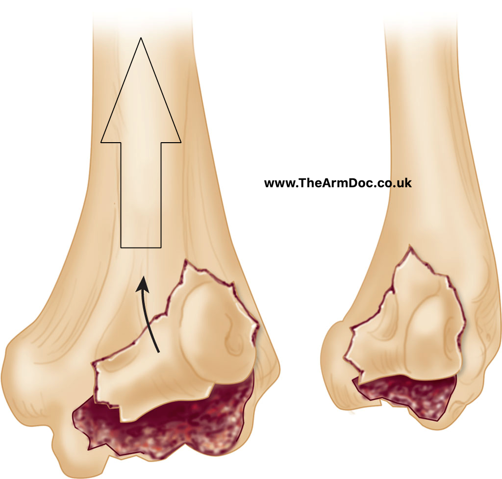
- Comminution: This means the bone is broken into several pieces, making the fracture complex.
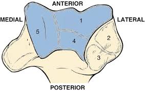
- Complex Bone Structure: The intricate shape of the distal humerus (the lower part of the upper arm bone) adds to the challenge of treating these fractures.
Initial Evaluation Goals
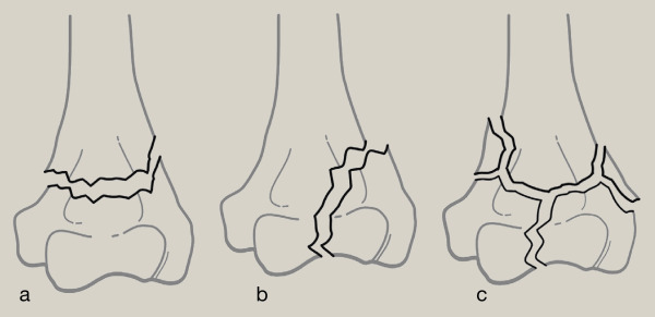
- Identify the Fracture Pattern: Understand how the bone is broken.
- Check for Previous Elbow Problems: Look for any existing elbow issues that might affect treatment.
- Assess Soft Tissue Damage: Determine if open wounds or significant soft tissue injuries exist.
- Identify Other Injuries: Look for related injuries in the arm or nearby nerves and blood vessels.
Diagnostic Imaging
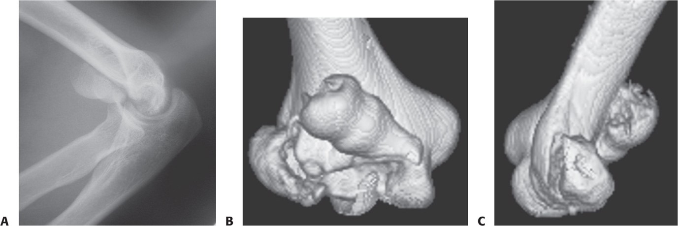
- X-rays: First step to visualise the fracture. Images are taken from the front (anteroposterior) and side (lateral) views.
- X-rays help identify fracture lines, fragments, and associated injuries in nearby bones.
- Due to the complexity of the distal humerus, X-rays might need to show the full extent of the fracture clearly.
- CT Scans with 3D Reconstruction: Provides a detailed view of the fracture, helping the surgeon plan the surgery more accurately.
- Shows the exact configuration of broken bone pieces and their positions
- Traction Radiographs: Taken in the operating room under anaesthesia to get a clearer picture if CT scans are unavailable.
Surgical Treatment
Internal Fixation
- Preferred Treatment: Most distal humerus fractures are treated with internal fixation.
- Modern Techniques: Include strategies to improve stability using pre-shaped plates and screws that lock into the plates.
- Elbow Replacement: This is considered for elderly patients with pre-existing elbow problems or very complex fractures due to weak bones.
- Goal: Achieve a stable bone construct that allows immediate movement without the risk of the fracture moving out of place again.
Fixation Techniques
-
Use of Plates and Screws:
- Plates are applied on both sides of the humerus to maximise fixation.
- Screws lock into these plates to provide additional stability.
-
Surgical Example:
-
- Parallel Plate Fixation: Two plates are placed on the humerus's medial (inner) and lateral (outer) sides.
- Post-surgery X-ray: This shows the bone pieces perfectly aligned and securely fixed using plates, with a clear picture of any additional repairs,'like fixing an olecranon osteotomy (a cut made in the elbow bone to allow better access during surgery).