“Advances in Minimally Invasive Techniques for Spine Surgery: Benefits and Procedures”

Rephrased Paragraph:

Traditional spine surgery has generally involved “open surgery,” requiring a long incision for the surgeon to fully view and access the spinal area. However, advancements in technology now allow many spine conditions to be managed through minimally invasive surgical techniques (MISS). These techniques achieve similar outcomes to open surgery but involve significantly smaller incisions, which translates to fewer muscle disruptions around the spine.

Compared to the larger incisions in open surgery, minimally invasive spine surgery reduces muscle damage, leading to less postoperative pain and a shorter recovery time. Despite these benefits, MISS can be technically demanding due to the limited access provided by smaller openings, requiring a more specialized skill set for surgeons. The criteria for opting for minimally invasive surgery are the same as those for open procedures.

Surgical intervention is generally considered only after nonsurgical treatments, such as medications and physical therapy, fail to alleviate symptoms. Additionally, a precise diagnosis, like a herniated disk or spinal stenosis, must be confirmed before recommending surgery.

Several minimally invasive techniques exist, all characterized by their use of smaller incisions and reduced muscle impact. These methods are commonly used for lumbar decompression and spinal fusion procedures. Decompression aims to alleviate nerve pressure by removing parts of a herniated disk or bone, while spinal fusion stabilizes painful vertebrae by fusing them into a single solid bone. This article provides an overview of minimally invasive approaches to spinal decompression and fusion.

For an in-depth guide on spinal fusion, covering methods, bone grafting, potential complications, and rehabilitation, see our complete overview on Spinal Fusion.

Description

Minimally invasive spine surgery (MISS), sometimes referred to as “less invasive spine surgery,” involves accessing the spine through small incisions with specialized instruments. In contrast to traditional open surgery, which requires a 5- to 6-inch incision, MISS minimizes tissue disruption by eliminating the need to pull muscles aside to view the spine.

In open spine surgery, retracting muscles allows the surgeon to:

  • Access the spine to remove damaged bone or intervertebral disks.
  • Clearly identify placement points for screws, cages, and bone graft materials to stabilize the spine and aid in healing.

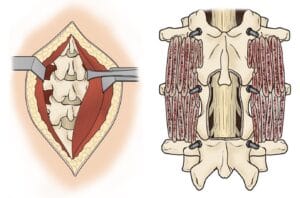
(Left) In traditional open surgery, muscles surrounding the spine are pulled back to reveal the bones. (Right) After removing portions of the bone (a decompression procedure called laminectomy), bone graft material and screws are placed along the sides of the vertebrae. 


However, open surgery often results in significant muscle and soft tissue damage due to muscle retraction, which may lead to new types of pain and an extended recovery time. The larger incision, along with tissue trauma, can increase blood loss and the risk of infection.

MISS was developed to address spinal issues with minimal harm to muscles and other tissues, offering a more targeted approach to the affected spine area. Key benefits of MISS include:

  • Smaller incisions
  • Reduced bleeding
  • Shorter or potentially no hospital stay, as some procedures can be done on an outpatient basis.


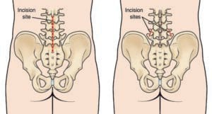
(Left) The incision site in the lower back that is used for some traditional open spine surgeries. (Right) The minimally invasive incisions typically used for a lumbar spinal fusion. Both the decompression and the placing of screws and rods are accomplished through these small incisions.

Procedure

MISS techniques, including fusion and decompression procedures like diskectomy and laminectomy, are performed using various methods, with the tubular retractor approach being the most common.

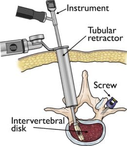
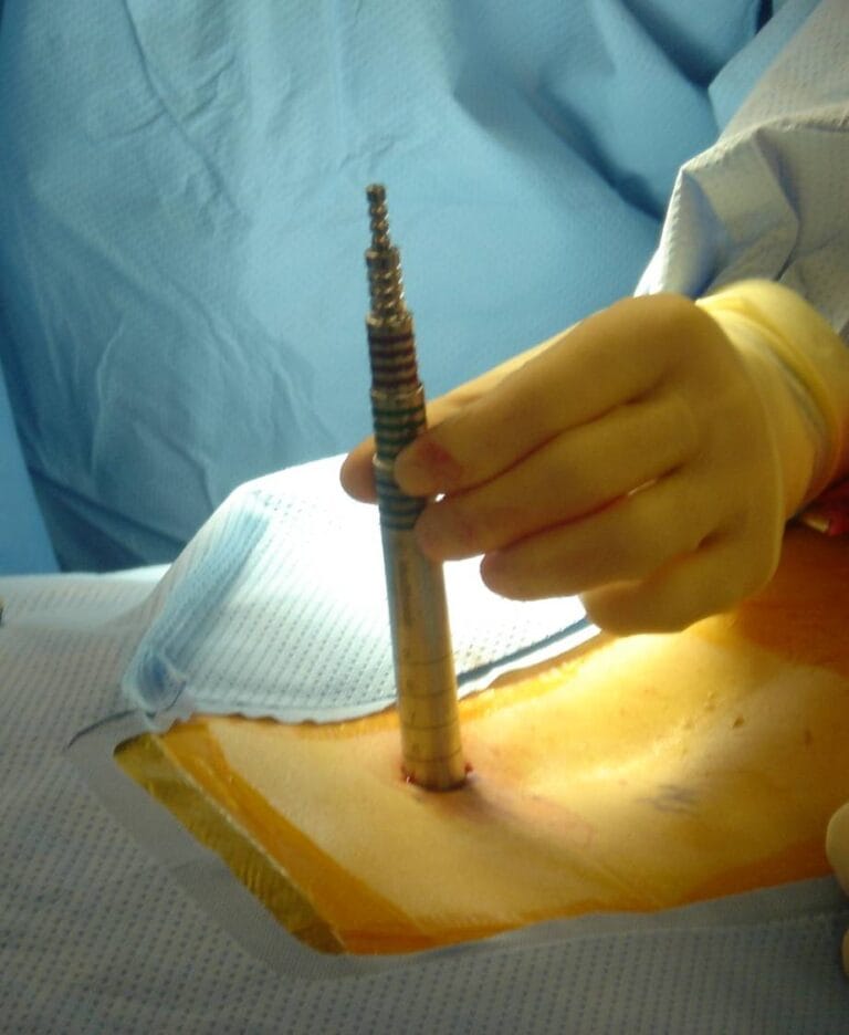
To begin, the surgeon makes a small incision and guides the tubular retractor through the skin and tissues down to the spinal area of concern. This creates a pathway directly to the target area without cutting the muscles, as the retractor keeps the tissues held open. Throughout the procedure, the surgeon uses small instruments that fit within the tubular retractor to access the spine, remove any bone or disk material, and insert any devices needed for fusion, such as screws or rods, through the same small incision. Some surgeries may require multiple retractors or incisions.

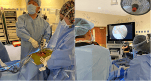
Fluoroscopy, a real-time X-ray imaging technique, is used to accurately position the incision and retractor, providing the surgeon with live images of the spine on a screen. A microscope often provides a magnified view of critical spine structures during surgery. Once the surgery is complete, the tubular retractor is removed, allowing the muscles to return to their natural position, significantly reducing the muscle damage typically seen in open procedures.


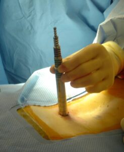
A tubular retractor is used to create a passageway for the surgeon to reach the problem area of the lower back.
Reproduced from Anderson D, Tannoury C: Minimally invasive lumbar surgery, Orthopaedic Knowledge Online Journal 2007; 5(7). Accessed January 2018. 


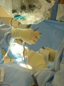
An operating microscope provides excellent illumination and magnification during minimally invasive spine procedures.
Reproduced from Anderson D, Tannoury C: Minimally invasive lumbar surgery, Orthopaedic Knowledge Online Journal 2007; 5(7). Accessed January 2018. 

Endoscopic Advances in Minimally Invasive Spine Surgery

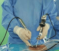
Innovative techniques in minimally invasive spine surgery continue to advance, with some surgeons now utilizing endoscopy—a small, high-definition camera similar to those used in knee and shoulder surgeries—to precisely access and treat spinal issues.

During an endoscopic spine procedure, the surgeon makes either a single 1 cm incision or two 7 mm incisions to reach the spine. A water-based system enhances visualization, allowing the surgeon to clearly see and address the affected area under high magnification. This approach is now being used for various procedures, including laminectomies, diskectomies, and even spinal fusions, broadening the potential applications of minimally invasive spinal surgery.


Endoscopic minimally invasive spine surgery.


During endoscopic minimally invasive spine surgery, water is used to provide better visualization of the spine, enabling surgeon to directly see and address the problem under greater magnification.


Anesthesia Options for Minimally Invasive Spine Surgery

Two primary types of anesthesia are commonly used in minimally invasive spine surgery (MISS):

  • General Anesthesia: You are fully asleep for the entire procedure.
  • Regional Anesthesia: You may remain awake, but sensation is entirely blocked from the waist down.

These options are selected based on the specific procedure and patient needs to ensure maximum comfort and safety during surgery.


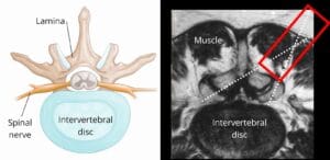
(Left) Cross-section view of a healthy intervertebral disk. (Right) Cross-section MRI scan showing the anatomy surrounding an intervertebral disk. The red rectangle shows placement of the tubular retractor through the muscle. The dotted lines show the positioning of small surgical instruments.

Common Minimally Invasive Spine Surgeries

MIS Lumbar Diskectomy

When a herniated disk in the lower back compresses a nerve, it can lead to significant leg pain, numbness, or weakness. To relieve these symptoms, the damaged portion of the disk is removed in a procedure called a diskectomy.

In an MIS lumbar diskectomy:

  • The patient is positioned face-down, and the surgeon makes a small incision over the herniated disk.
  • A retractor is inserted, and a small part of the lamina bone is removed to give the surgeon a view of the spinal nerve and the affected disk.
  • The nerve is gently retracted, and only the damaged part of the disk is carefully removed.

This minimally invasive method is also applicable to herniated disks in the neck. In this case, the surgery is performed through the back of the neck and is known as a MIS posterior cervical foraminotomy/diskectomy.


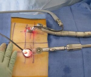
In an MIS TLIF procedure, a tubular retractor is placed on either side of the spine.

A small instrument is inserted through the tubular retractor to remove a herniated disk. Screws to support the fusion are placed in the bone through both retractors.

MIS Lumbar Fusion

A traditional open lumbar fusion can be performed from various approaches—through the back, abdomen, or side. Minimally invasive lumbar fusion techniques provide similar access while reducing tissue disruption.

One common approach in MISS is the transforaminal lumbar interbody fusion (TLIF), where the surgeon accesses the spine slightly off-center. This approach minimizes nerve manipulation.

In an MIS TLIF:

  • The patient is placed face-down, and retractors are positioned on either side of the spine, preserving the midline ligaments and bone.
  • Using the retractors, the surgeon removes the lamina and disk material, places a bone graft into the disk space, and stabilizes the area with screws or rods.
  • In some cases, additional bone graft materials are used to enhance healing potential.

Lateral fusion approaches, such as extreme lateral interbody fusion (XLIF) and direct lateral interbody fusion (DLIF), avoid injury to the back muscles and do not require manipulation of spinal nerves. A newer variation, oblique lateral interbody fusion (OLIF), also makes an incision on the side but approaches the spine at an angle to preserve the psoas muscle, achieving similar outcomes to the other lateral methods.

Complications

As with any surgery, MISS carries some risks, though studies suggest that infection rates may be lower with MISS compared to open spinal fusion procedures. Before surgery, your doctor will discuss potential risks and take steps to minimize them.

Possible complications of MISS include:

  • Infection: Patients receive antibiotics before, during, and after surgery to reduce infection risk.
  • Bleeding: Some blood loss is expected, but it’s usually minimal.
  • Pain at the Graft Site: A small number of patients may experience prolonged discomfort at the bone graft site.
  • Recurring Symptoms: Some patients might experience a return of initial symptoms.
  • Pseudarthrosis: In this condition, the bone does not fully heal, potentially requiring additional surgery. Smoking increases the risk of pseudarthrosis.
  • Nerve Damage: Rarely, nerves or blood vessels may be affected.
  • Blood Clots: Blood clots in the legs (deep vein thrombosis) are rare but can be dangerous if they travel to the lungs.

Recovery

MISS generally leads to shorter hospital stays, with some patients able to go home the same day or within 1 to 2 days. In contrast, traditional spine surgery often requires a 3- to 5-day hospital stay. Since minimally invasive methods limit muscle and tissue disruption, post-operative pain is typically reduced compared to open surgeries. Although some discomfort is expected, advancements in pain management can make recovery more comfortable.

To help rebuild strength and facilitate recovery, your doctor may recommend physical therapy, depending on your procedure and physical condition. Specific exercises will assist you in regaining strength for daily activities and work.

For patients who undergo spinal fusion, the bone may take several months to solidify, although pain relief is often achieved sooner. During this healing period, proper alignment is essential, and you’ll receive guidance on safe ways to move, sit, stand, and walk.

The timeline for returning to daily activities after MISS varies by procedure and individual factors. Your doctor will evaluate your progress post-surgery to ensure a successful recovery.

Scroll to Top