Understanding Lumbar Spinal Canal Narrowing: Causes, Symptoms, and Age-Related Risk Factors

Lumbar spinal canal narrowing is a frequent source of lower back pain and leg discomfort, often leading to sciatica symptoms.

As the body ages, natural wear-and-tear can gradually reduce the width of the spinal canal, which encases the spinal nerves and spinal cord—a condition known as spinal stenosis. Degenerative spinal changes are common, appearing in as many as 95% of individuals by age 50, and lumbar spinal stenosis predominantly affects adults over 60. Both men and women experience similar rates of nerve root compression.

In rare cases, some individuals are born with spinal structures that predispose them to lumbar spinal stenosis, referred to as congenital spinal stenosis. This typically occurs in people with a naturally smaller spinal canal, where restricted space makes them more susceptible to early degeneration or arthritis. This form of stenosis is more common in men, with symptoms generally beginning between ages 30 and 50.

Anatomy

The spine consists of a series of small bones, called vertebrae, stacked along the back to form a sturdy column. Alongside these vertebrae, the spine comprises muscles, ligaments, nerves, and intervertebral discs, all essential for stability, flexibility, and protection.

A basic understanding of spine structure and function is key to grasping spinal stenosis. For an overview of spine anatomy, visit our section on Spine Basics.

Description

Spinal stenosis develops when the space surrounding the spinal cord and nerves becomes restricted. This narrowing can place pressure on the spinal cord and nerve roots, often resulting in pain, numbness, or leg weakness.

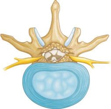
 

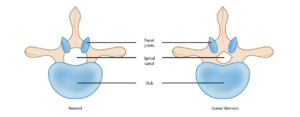
This illustration shows a healthy vertebra (cross-section view) and a vertebra with narrowing of the spinal canal, called stenosis.

Cause

The primary cause of spinal stenosis is arthritis, which involves the degeneration of joints throughout the body.

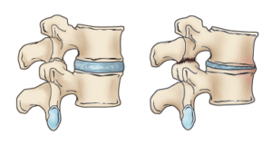
In the spine, arthritis can develop as intervertebral discs deteriorate and lose moisture. Discs in children and young adults are typically rich in water, maintaining their flexibility and cushioning abilities. However, as we age, these discs gradually dry out and weaken. This degeneration can lead to disc settling or collapse, resulting in reduced disc height and contributing to spinal canal narrowing.


When we are young, disks have a high water content (left). As disks age and dry out, they may lose height or collapse (right). This puts pressure on the facet joints and may result in arthritis


In youth, intervertebral discs are rich in water content (left), which helps them maintain height and resilience. As we age, these discs gradually lose moisture, potentially reducing in height or even collapsing (right). This shift increases pressure on the facet joints, which can lead to the development of arthritis.

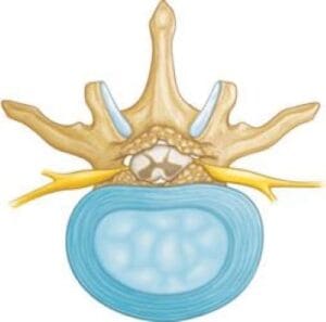
Arthritic bone spurs narrow the spinal canal.

Symptoms

  1. Back Pain: Individuals with spinal stenosis may experience back pain, though the presence and intensity can vary based on the severity of arthritis in the spine.
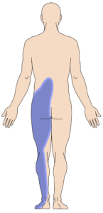
  2. Burning Pain in Buttocks or Legs (Sciatica): Nerve compression in the spine may lead to pain in the areas supplied by these nerves, often described as an aching or burning sensation. Typically, the pain originates in the buttocks and radiates down the leg, and in advanced cases, it can extend to the foot.
  3. Numbness or Tingling in Buttocks or Legs: Increased nerve pressure may cause numbness and tingling alongside the burning pain, although not everyone will experience both symptoms simultaneously.
  4. Leg Weakness or Foot Drop: When nerve compression reaches a significant level, weakness may develop in one or both legs. Some people may experience foot drop, where lifting the front of the foot becomes difficult, resulting in the sensation that the foot “slaps” against the ground while walking.
  5. Acute Cauda Equina Syndrome: This uncommon but serious condition is a medical emergency requiring immediate attention. Severe nerve compression can cause numbness in the groin area, loss of bowel or bladder control, and leg weakness that impedes walking. These symptoms indicate the need for emergency surgery.

In spinal stenosis, leaning forward or sitting often relieves discomfort, as studies suggest that these positions can expand the space available for nerves in the lumbar spine. Pain generally intensifies when standing straight or walking. Some individuals find they can comfortably use a stationary bike or walk with support, like leaning on a shopping cart. However, walking for more than 1 or 2 blocks may provoke severe sciatica or leg weakness.


Spinal nerves relay sensation in specific parts of your body. Pressure on the nerves can cause pain in the areas that the nerves supply, including pain in the buttocks that radiates down the leg — called sciatica.

Doctor Examination

Medical History and Physical Examination
During the initial examination, your doctor will review your symptoms and medical history. They will assess your back by examining it closely and applying pressure to different areas to identify any painful spots. You may be asked to bend forward, backward, and side-to-side to help determine any movement limitations or pain.

Imaging Tests

To confirm a diagnosis, your doctor may order imaging tests:

  • X-rays: While X-rays primarily capture bone images, they can reveal signs of spinal stenosis, such as reduced disc height or bone spurs. X-rays taken during forward and backward bending can also help detect joint instability and excessive spinal mobility, a condition known as spondylolisthesis.
  • Magnetic Resonance Imaging (MRI): An MRI provides detailed images of soft tissues like muscles, discs, nerves, and the spinal cord, offering a clearer view than X-rays.
  • Additional Tests: Computed tomography (CT) scans produce cross-sectional images of the spine, providing a more detailed view of bone structures. A myelogram, involving the injection of dye into the spine, can also help highlight compressed nerves.

Treatment

Nonsurgical Treatment

Nonsurgical options aim to alleviate pain and improve function, even though they do not reverse the spinal canal narrowing. Many individuals find relief through these methods:

  • Physical Therapy: Stretching exercises, massage, and strengthening exercises for the lumbar and abdominal muscles can help manage symptoms.
  • Lumbar Traction: While beneficial for some, traction provides limited results, with minimal scientific evidence supporting its effectiveness.
  • Anti-inflammatory Medications: Pain associated with spinal stenosis is often due to inflammation around the spinal nerves. Non-steroidal anti-inflammatory drugs (NSAIDs) help relieve pain and, over a course of 5-10 days, can offer added anti-inflammatory benefits. Both over-the-counter and prescription-strength NSAIDs are available, but they should be used cautiously to avoid side effects like gastritis, ulcers, and kidney issues.
  • Steroid Injections (Epidural): Cortisone, a powerful anti-inflammatory, can be injected around the nerves or into the epidural space to reduce swelling and pain, although it does not relieve weakness in the legs. No more than three injections per year are recommended.
  • Acupuncture: This method can relieve pain in mild cases of lumbar stenosis. While safe, its long-term effectiveness has not been scientifically validated.
  • Chiropractic Manipulation: Chiropractic adjustments may alleviate some lumbar stenosis pain, though those with osteoporosis or a herniated disc should exercise caution, as spinal manipulation may worsen symptoms or cause additional injuries.

Surgical Treatment

Surgery is generally considered when pain and weakness severely impact quality of life, especially with walking difficulties. The main surgical options for lumbar stenosis are laminectomy and spinal fusion, both of which have shown to effectively reduce pain. Discuss the pros and cons of each with your doctor.

  • Laminectomy: This decompression procedure involves removing bone, bone spurs, and ligaments that press on the nerves. It can be done as open surgery, requiring a single incision, or through a minimally invasive approach with several smaller incisions.
  • Spinal Fusion: When arthritis causes spinal instability, spinal fusion is often combined with decompression to stabilize the spine.

Rehabilitation

After surgery, your hospital stay will depend on your health and the procedure performed. Patients undergoing decompression may go home the same day or the next and return to normal activities within a few weeks. Fusion surgery typically requires an additional 2-3 days of hospitalization.

Your doctor may prescribe a brace or corset for comfort and encourage you to begin walking early on. Most patients require physical therapy to strengthen their back muscles. Your physical therapist will guide you through exercises to build core strength, endurance, and flexibility for spinal stability, creating a personalized program based on your health and history.

Most people with desk jobs can return to work within days to weeks post-surgery and resume regular activities within 2-3 months. Older adults requiring more care may be transferred to a rehabilitation facility before returning home.

Postoperative Opioid Medication: Patients new to opioid use may require a short course of opioids following laminectomy, while those undergoing fusion may need them for a longer period. Long-term users may continue opioids post-surgery due to established dependency.

Surgical Risks

All surgeries carry minor risks, including bleeding, infection, blood clots, and anesthesia reactions. People who are elderly, overweight, diabetic, smokers, or have multiple health conditions may face higher risks of complications.

Specific risks of spinal stenosis surgery include:

  • Dural tear (a tear in the sac covering the nerves)
  • Failed bone fusion (if fusion was performed)
  • Failure of fusion hardware (screws or rods)
  • Nerve damage
  • Possible need for additional surgery
  • Incomplete symptom relief or recurrence of symptoms

Surgical Outcomes

Laminectomy, with or without spinal fusion, generally yields good to excellent outcomes for most patients with lumbar stenosis. Leg pain typically improves more than back pain, allowing many individuals to resume an active lifestyle following recovery.


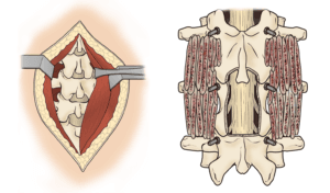
(Left) In traditional open surgery, to access the spine, the surgeon pulls back muscles to expose the bone.  (Right) After the laminectomy, the surgeon places bone graft material and screws along the sides of the vertebrae to help with healing. 

Scroll to Top