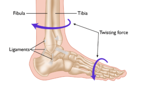
Pediatric ankle fractures, commonly referred to as broken ankles, are among the most frequent injuries in children. These fractures involve a break in one or more bones of the ankle joint: the tibia (shinbone), the fibula (outer ankle bone), or the talus (a small bone located at the back of the foot).

The severity of ankle fractures in children can vary widely. Minor fractures are often effectively treated with a simple walking boot, while complex fractures may necessitate surgical intervention. A pediatric orthopedic specialist will guide you through the available treatment options and provide ongoing follow-up care to ensure proper healing and monitor long-term recovery outcomes.

Anatomy of the Ankle Joint

The ankle joint is the point where the tibia (shinbone), fibula (outer ankle bone), and talus (a small bone in the foot) come together. These bones are held in place by ligaments, which function like sturdy ropes, providing stability to the joint by connecting bones and preventing excessive movement. The ankle contains several important ligaments that work together to maintain joint stability and facilitate smooth movement.
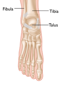
 

The normal skeletal anatomy of the foot and ankle.

Understanding Ankle Fractures in Children

In children and adolescents, ankle fractures typically affect the tibia or fibula, often involving the growth plates—specialized areas of cartilage near the ends of long bones. Growth plates play a critical role in bone development, gradually hardening into solid bone as the child matures. However, because these growth plates are the last parts of the bones to solidify, they are particularly prone to injury and fractures.

As adolescents approach the end of their growth phase, growth plates begin to close and harden, making them susceptible to transitional fractures during this maturing stage. Two common types of transitional ankle fractures are triplane fractures and Tillaux fractures, which require specific treatment approaches depending on their severity and location (refer to the Treatment section for more details)
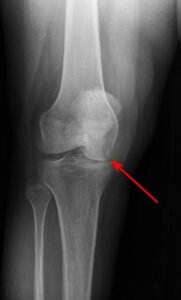
This X-ray of a child’s ankle taken from the front clearly shows the growth plates of the tibia and fibula (red arrows).

Causes of Pediatric Ankle Injuries

Ankle injuries in children often result from sudden twisting of the lower leg or foot, frequently occurring during sports or active play. High-energy activities, especially those involving lateral movements and jumping, significantly increase the risk of such injuries.

Sports like basketball, for instance, pose a higher risk because players often jump to defend, shoot, or rebound. If a child lands awkwardly or on another player’s foot, it can cause the ankle to twist or roll inward or outward, leading to potential fractures or sprains. Recognizing these risks can help in adopting preventative measures to minimize injury during physical activities.

A twisting force to the lower leg or foot is a common cause of ankle fractures, as well as ligament injuries (sprains).

Symptoms of Pediatric Ankle Fractures

Differentiating between an ankle sprain and a fracture can be challenging, as both conditions initially cause pain and swelling. However, fractures often present additional signs and symptoms, such as:

  • Inability to stand or walk
  • Bruising
  • Tenderness to the touch
  • Visible deformity in the ankle area

Doctor Examination

Medical History and Physical Examination

A thorough evaluation begins with the doctor discussing your child’s medical history and the circumstances of the injury. This is followed by a careful physical examination of the ankle to assess:

  • Swelling and bruising
  • Tenderness at the injury site
  • Any deformity or misalignment of the ankle
  • Presence of tears or open wounds in the skin
  • Proper functioning of arteries and nerves to rule out further complications

Imaging Tests

When a fracture is suspected, imaging tests are crucial for an accurate diagnosis:

  • X-rays: The most commonly used tool for diagnosing fractures, X-rays provide clear images of the bone structure, helping determine whether a bone is broken.
  • Computed tomography (CT): A CT scan creates detailed cross-sectional images of the ankle bones, which are especially helpful for fractures that extend into the ankle joint or require surgical intervention.
  • Magnetic resonance imaging (MRI): In cases where a fracture is suspected but not visible on X-rays, an MRI may be ordered. MRI scans provide high-resolution images of both bones and soft tissues, such as ligaments, offering a comprehensive view of the injury.

These diagnostic steps ensure that your child’s condition is thoroughly evaluated, enabling precise treatment and a better recovery outcome.

Scroll to Top