Introduction
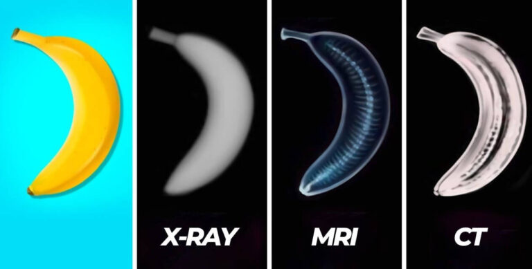
Medical imaging plays a crucial role in diagnosing and monitoring various health conditions. Among the most commonly used imaging techniques are X-rays, Computed Tomography (CT) scans, and Magnetic Resonance Imaging (MRI). While all three techniques provide detailed internal images of the body, they operate on different principles and are suitable for different medical conditions.
This article provides a comprehensive analysis of these imaging modalities, including their working mechanisms, advantages, limitations, applications, risks, and comparisons. It also adheres to SEO best practices to improve visibility in search engines.
1. X-rays
1.1 What Are X-rays?
X-rays are a type of electromagnetic radiation with high energy and short wavelength. They are widely used in medicine for visualizing bones, detecting fractures, and diagnosing lung conditions.
1.2 How Do X-rays Work?
X-ray machines emit ionizing radiation that passes through the body. Different tissues absorb radiation at different rates:
- Bones absorb most of the X-rays, appearing white.
- Soft tissues (muscles, organs, fat) allow more X-rays to pass, appearing in shades of gray.
- Air-filled areas (lungs) absorb very little X-ray, appearing black.
1.3 Features of X-ray Imaging
- Quick and widely available.
- Relatively low cost compared to CT or MRI.
- High contrast for bones and dense structures.
- Non-invasive but involves ionizing radiation.
1.4 Applications of X-rays
- Skeletal system: Fractures, arthritis, bone tumors.
- Respiratory system: Pneumonia, lung infections, tuberculosis.
- Dental care: Tooth decay, jaw disorders.
- Mammography: Detecting breast cancer.
- Abdominal imaging: Detecting foreign objects, bowel obstruction.
1.5 Limitations of X-rays
- Limited soft tissue contrast: Cannot effectively distinguish muscles, tendons, or internal organs.
- Radiation exposure: Prolonged or repeated exposure increases health risks.
2. CT Scans (Computed Tomography)
2.1 What Is a CT Scan?
A Computed Tomography (CT) scan is an advanced imaging technique that uses X-rays and computer processing to create cross-sectional images of the body.
2.2 How Does a CT Scan Work?
A CT scanner rotates around the body, taking multiple X-ray images from different angles. A computer reconstructs these images into a detailed 3D visualization.
2.3 Features of CT Scanning
- Provides detailed images of bones, organs, and blood vessels.
- Higher resolution than standard X-rays.
- Quick scan time (typically a few minutes).
- Can be enhanced with contrast dyes for better visibility.
2.4 Applications of CT Scans
- Head and brain: Stroke, brain tumors, skull fractures.
- Chest and lungs: Pulmonary embolism, lung nodules, COVID-19 complications.
- Abdomen and pelvis: Appendicitis, kidney stones, liver disease.
- Cardiology: Coronary artery disease, aneurysms.
- Trauma and emergencies: Internal bleeding, organ damage.
2.5 Limitations of CT Scans
- Higher radiation exposure than X-rays.
- Contrast agents may cause allergic reactions.
- Not suitable for pregnant women due to radiation risks.
3. MRI (Magnetic Resonance Imaging)
3.1 What Is an MRI?
MRI is a non-invasive imaging technique that uses strong magnetic fields and radio waves to produce detailed images of soft tissues, organs, and joints.
3.2 How Does MRI Work?
MRI scanners create a strong magnetic field that aligns hydrogen atoms in the body. Radiofrequency pulses then cause these atoms to emit signals, which a computer processes into detailed images.
3.3 Features of MRI Scanning
- Superior soft tissue contrast compared to X-rays and CT.
- No radiation exposure (safe for repeated use).
- Excellent for imaging the brain, spinal cord, joints, and soft tissues.
3.4 Applications of MRI
- Neurology: Brain tumors, multiple sclerosis, stroke.
- Orthopedics: Ligament tears, cartilage damage, herniated discs.
- Cardiology: Heart abnormalities, congenital defects.
- Abdominal imaging: Liver, pancreas, kidneys.
- Oncology: Detecting and monitoring tumors.
3.5 Limitations of MRI
- Longer scan time (30-60 minutes).
- Loud noise and confined space may cause discomfort.
- Not suitable for patients with metal implants.
- More expensive than X-rays and CT scans.
4. Comparison of X-ray, CT, and MRI
4.1 Imaging Capabilities
| Feature | X-ray | CT Scan | MRI |
|---|---|---|---|
| Best for Bones | ✅ Excellent | ✅ Very Good | ❌ Limited |
| Soft Tissue Contrast | ❌ Poor | ✅ Moderate | ✅✅ Excellent |
| Radiation Exposure | ✅ Low | ❌ High | ✅✅ None |
| Speed of Scan | ✅ Very Fast | ✅ Fast | ❌ Slow |
| Cost | ✅ Low | ✅ Moderate | ❌ High |
4.2 When to Use Which Imaging Modality?
- Use X-rays for bone fractures, lung conditions, and dental issues.
- Use CT scans for detailed 3D imaging of organs, head trauma, and emergency diagnostics.
- Use MRI for brain imaging, spinal cord injuries, and soft tissue evaluation.
5. Safety Considerations
5.1 Radiation Risks
- X-rays and CT scans use ionizing radiation, which can cause cell damage in high doses.
- MRI does not use radiation, making it a safer option for repeated scans.
5.2 Contrast Agents
- CT and MRI scans often require contrast agents (iodine-based for CT, gadolinium-based for MRI).
- Patients with kidney disease may be at risk for complications from contrast dyes.
5.3 MRI Safety Concerns
- Metal implants (pacemakers, clips, implants) can be dangerous in an MRI due to strong magnetic fields.
- Claustrophobic patients may struggle with MRI scans due to the enclosed space.
6. Conclusion
Each imaging technique—X-ray, CT scan, and MRI—has its unique advantages and limitations. Understanding when to use each modality is crucial for accurate diagnosis and minimizing unnecessary risks.
Summary of Recommendations:
- X-ray: Best for bones and lungs.
- CT scan: Best for organs, emergencies, and detailed internal imaging.
- MRI: Best for soft tissues, nerves, and the brain.