Understanding Transient Hip Bone Loss: Causes, Symptoms, and Recovery

Transient osteoporosis of the hip is an uncommon condition characterized by temporary bone loss in the upper section of the thighbone (femur).

Individuals with this condition often experience a sudden onset of hip pain, which intensifies during walking or weight-bearing activities. Over time, this discomfort can escalate, potentially becoming severe enough to interfere with daily activities.

Fortunately, the painful symptoms usually subside gradually and disappear entirely within 6 to 12 months. In most cases, the affected hip regains its normal bone strength during the recovery process.

It’s important to note that transient osteoporosis of the hip is distinct from age-related osteoporosis. Unlike the progressive, painless bone weakening associated with age-related osteoporosis—which increases the risk of fractures throughout the body—transient osteoporosis is temporary and localized, with a defined recovery period.

Anatomy

The hip is one of the largest and most crucial joints in the human body, functioning as a ball-and-socket joint. The socket, known as the acetabulum, is part of the pelvic bone, while the ball is the femoral head, which forms the upper portion of the femur (thighbone). This structure allows for a wide range of motion and supports the body’s weight during activities such as walking and running.
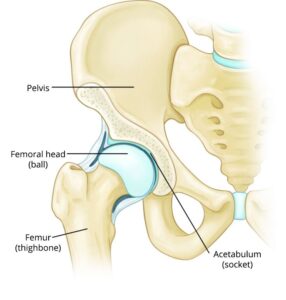
 

The hip is a ball-and-socket joint. In a healthy hip, the femoral head (ball) fits firmly into the acetabulum (socket).

Description

Transient osteoporosis of the hip occurs when the femoral head temporarily weakens and experiences a loss of bone density (bone mass). During this period of reduced bone strength, the risk of fractures increases significantly.

Although this condition most commonly affects the hip joint, it can also occur in other joints of the lower extremities, such as the knee, ankle, or foot.

Transient osteoporosis of the hip predominantly affects young to middle-aged men (typically between 30 and 60 years old). It is also more common among women in the final trimester of pregnancy or those who have recently given birth. Research published in the Journal of Obstetrics and Gynecology in 2017 suggests that approximately 1 in 250,000 pregnant women experience this condition.

Related Articles

  • Staying Healthy: Bone Health Basics
  • Recovery: How To Use Crutches, Canes, and Walkers
  • Diseases & Conditions: Osteoporosis
  • Staying Healthy: Preventing Back Pain: Tips for New Parents

Cause

The exact cause of transient osteoporosis of the hip remains unknown. While researchers are actively investigating, no definitive explanation has been established. Several potential causes have been proposed, including:

  • Obstruction of small blood vessels surrounding the hip
  • Hormonal changes, particularly during pregnancy or postpartum
  • Abnormal mechanical stresses, such as excessive force or load on the bone

Symptoms

The hallmark symptoms of transient osteoporosis of the hip include:

  • A sudden onset of pain localized in the front of the thigh, groin, hip, or buttocks
  • Pain that worsens with weight-bearing activities but may ease with rest
  • No history of trauma or injury to the hip that could account for the pain
  • Slightly reduced range of motion; mild movements are typically painless, but extreme hip motions may trigger discomfort
  • Progressive pain that intensifies over several weeks or months and can become debilitating
  • A noticeable limp as a response to protect the joint and minimize pain

These symptoms often develop gradually, making early diagnosis crucial to managing the condition and preventing further complications.

Doctor Examination: What to Expect

Medical History and Physical Assessment

During your medical consultation, your doctor will review your symptoms and discuss when they first began. They may inquire if you recall any specific injury or trauma to your hip that could explain the discomfort.

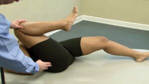
As part of the physical examination, your doctor will assess your hip’s range of motion by guiding your leg through various movements. This helps evaluate how the joint responds to motion and pinpoints the source of the pain.

Patients with transient osteoporosis of the hip typically report greater discomfort when they actively move their hip (active range of motion) compared to when the doctor moves it for them (passive range of motion). Additionally, pain is often most noticeable during extreme hip movements or weight-bearing activities.

This distinction—severe pain during weight-bearing but minimal discomfort with passive movements—is a key diagnostic indicator of transient osteoporosis of the hip. These findings, combined with your medical history, guide your doctor toward an accurate diagnosis and appropriate treatment plan.


During the exam, your doctor will test the range of motion in your hip.


Diagnostic Tests for Transient Osteoporosis of the Hip

X-rays

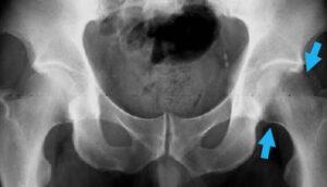
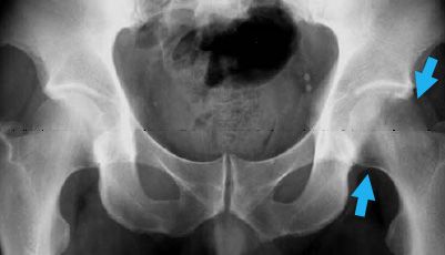
X-rays play a vital role in diagnosing transient osteoporosis of the hip, although the findings can vary depending on the stage of the condition. During the early stages (typically within the first 6 weeks), X-ray images may reveal a slight reduction in the bone density of the femoral head. However, these changes are often subtle and can be difficult to detect.

As the condition progresses over several months, X-rays may show a significant loss of bone density, with the femoral head appearing almost entirely diminished on the images. This temporary and reversible bone density loss is the defining characteristic of transient osteoporosis, giving the condition its name.

Early detection through imaging is crucial to monitor the disease’s progression and guide treatment strategies effectively.


This X-ray of the pelvis shows early changes in bone density in the affected hip (arrows).


Advanced Imaging and Diagnostic Tests for Transient Osteoporosis of the Hip

Other Imaging Scans

Since X-rays often fail to detect bone loss in the early stages of transient osteoporosis, doctors may recommend additional imaging tests for a more accurate diagnosis.

  • Computed Tomography (CT) and Magnetic Resonance Imaging (MRI):
    These imaging techniques provide detailed views of the hip joint. MRI scans, in particular, are effective for visualizing the surrounding soft tissues, including muscles, tendons, and ligaments, making them a valuable tool in diagnosing transient osteoporosis of the hip.
  • Nuclear Medicine Bone Scan:
    This imaging test is particularly useful for identifying bone changes caused by infection or injury. It’s important to note that a nuclear medicine bone scan is different from a dual-energy X-ray absorptiometry (DEXA) scan. While DEXA scans are the gold standard for detecting age-related osteoporosis, they are not effective for diagnosing transient osteoporosis of the hip.

If you are pregnant, your doctor may postpone certain imaging studies until the later stages of pregnancy or after delivery. However, MRI is generally considered safe during pregnancy. Discuss the risks and benefits with your orthopedic surgeon, primary care doctor, and obstetrician before undergoing this test.

Laboratory Tests

Although there is no specific blood test to diagnose transient osteoporosis of the hip, laboratory tests can help rule out other potential causes of hip pain. These may include metabolic disorders, endocrine conditions, or metastatic diseases (cancers).

Bone Marrow Edema

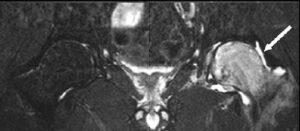
One of the hallmark signs of transient osteoporosis of the hip is bone marrow edema, which is characterized by inflammation and fluid accumulation in the bone marrow. This spongy tissue, located in the hollow parts of long bones, plays a crucial role in blood cell production.

MRI scans are particularly effective at identifying bone marrow edema and are among the most reliable imaging studies for diagnosing transient osteoporosis of the hip. The presence of bone marrow edema on an MRI scan often provides a definitive clue in confirming the condition.


This MRI image shows edema surrounding the affected hip. Edema causes the bone to appear white in the MRI image.

Treatment Options for Transient Osteoporosis of the Hip

Although transient osteoporosis of the hip resolves on its own, treatment is aimed at alleviating symptoms and protecting the bones from further damage during the period of weakened bone density.

1. Non-Steroidal Anti-Inflammatory Drugs (NSAIDs):
Medications like ibuprofen and naproxen are commonly prescribed to reduce pain and inflammation associated with the condition.

2. Weight-Bearing Restrictions:
Your doctor may recommend temporarily avoiding or significantly limiting weight-bearing activities to reduce stress on the affected hip. Using assistive devices such as crutches, a cane, or a walker can help minimize pressure on the hip and decrease the risk of fractures while the bone is weakened.

3. Physical Therapy:
To maintain muscle strength and flexibility around the hip, your doctor may suggest specific exercises, especially as pain begins to subside. Water-based exercises can be particularly beneficial because they promote movement while minimizing weight-bearing stress on the joint.

4. Nutrition:
Adequate nutrition plays a crucial role in the recovery process. Ensuring a diet rich in calcium and vitamin D can support bone healing and help restore bone density more effectively.

Expected Outcomes

With timely diagnosis and appropriate treatment, most individuals recover from transient osteoporosis of the hip within 6 to 12 months. In the majority of cases, bone strength in the hip returns to normal, and symptoms fully resolve.

However, a small percentage of patients may experience a recurrence of transient osteoporosis later in life. The condition may reappear in the same hip or develop in the opposite hip. Unfortunately, it is challenging to predict if or when the condition might return.

Scroll to Top