Femoroacetabular impingement (FAI) occurs when excess bone develops along one or both of the bones forming the hip joint, resulting in an uneven shape. This irregularity prevents the bones from aligning properly, causing them to make contact during movement. Over time, this repeated friction can lead to joint damage, manifesting as pain and restricted mobility.
Anatomy
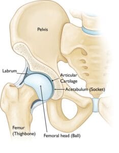
The hip functions as a ball-and-socket joint, where the socket, known as the acetabulum, is part of the large pelvic bone, and the ball is the femoral head, or the upper portion of the thighbone (femur). A smooth, slippery layer called articular cartilage coats both the ball and socket surfaces, reducing friction and allowing the bones to glide smoothly during movement. Surrounding the acetabulum is the labrum, a sturdy ring of fibrocartilage. This labrum acts like a gasket, sealing the joint securely and contributing to hip stability.
In a healthy hip, the femoral head fits perfectly into the acetabulum.
Description
In femoroacetabular impingement (FAI), the femoral head and/or acetabulum develop abnormal bony growths, or “bumps,” which can occur due to:
- A congenital shape of the hip bones
- Bone overgrowth or the formation of bone spurs
These extra bone formations disrupt the smooth movement of the hip joint, causing the bones to make abnormal contact. Over time, this can lead to labral tears and deterioration of the articular cartilage, potentially resulting in osteoarthritis.
Types of FAI
There are three primary types of FAI:
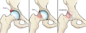
- Pincer Impingement: This type arises when additional bone extends beyond the acetabulum’s normal rim, compressing the labrum under the protruding rim.
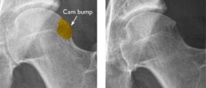
- Cam Impingement: In cam impingement, the femoral head is irregularly shaped and doesn’t rotate smoothly within the acetabulum. A bump on the femoral head’s edge grinds against the acetabular cartilage.
- Combined Impingement: This form involves both pincer and cam types, with features of each contributing to the impingement.
(Left) Pincer impingement. (Center) Cam impingement. (Right) Combined impingement.
Cause
Femoroacetabular impingement (FAI) develops due to abnormal hip bone formation during growth in childhood. This structural irregularity, involving a cam or pincer bone spur or both, ultimately contributes to joint damage and discomfort. When hip bones are shaped this way, preventive measures are limited, making FAI a challenge to avoid.
The prevalence of FAI remains uncertain, and while some individuals with FAI may lead active lives without issues, symptoms often indicate cartilage or labral damage and can signal a progression of the condition. Athletes and highly active individuals might experience symptoms earlier due to greater hip joint use, but physical activity itself does not cause FAI.
Symptoms
Common symptoms of FAI include:
- Pain
- Stiffness
- Limping
Pain is typically concentrated in the groin area but may also be felt on the outer hip. Movements such as turning, twisting, or squatting may trigger a sharp, stabbing sensation, though some individuals may experience a dull ache instead.
Home Remedies
When symptoms first appear, try identifying any activities that may have triggered the pain. Adjusting your movements and allowing your hip to rest can help alleviate discomfort. Over-the-counter anti-inflammatory medications, such as ibuprofen or naproxen, can also relieve pain.
If symptoms continue, consult a healthcare provider to pinpoint the cause of the pain and discuss treatment options. Ignoring persistent symptoms may lead to increased hip joint damage from FAI.
Doctor Examination
At your initial consultation, the doctor will review your overall health and assess your hip symptoms. A thorough hip examination will follow.
Impingement Test
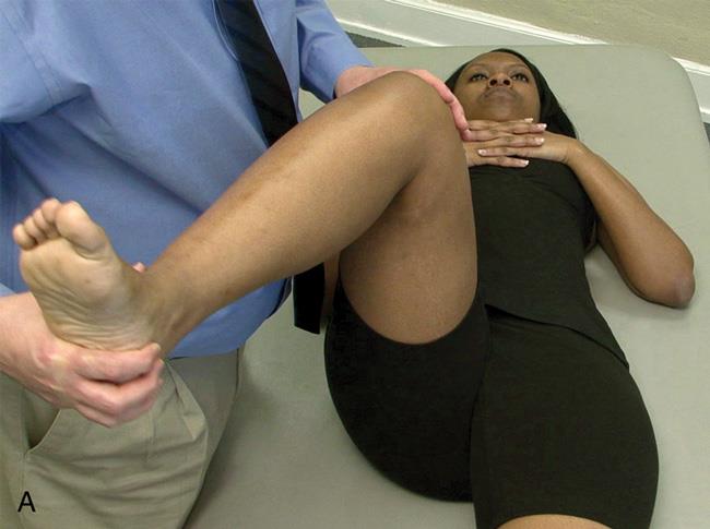
During the examination, your doctor may perform an impingement test. In this test, your doctor will bring your knee up toward your chest and rotate it inward toward the opposite shoulder. If this motion replicates your hip pain, it indicates a positive result for impingement.
Impingement test.
Imaging Tests
To accurately diagnose femoroacetabular impingement (FAI), your doctor may recommend a series of imaging tests.
- X-rays: X-rays provide detailed images of bone structure, allowing your doctor to identify the abnormal bone shapes characteristic of FAI. They can also reveal early signs of arthritis.
- Computerized Tomography (CT) Scans: CT scans offer a more precise view than standard X-rays, enabling your doctor to closely examine the specific irregularities in your hip’s shape.
- Magnetic Resonance Imaging (MRI) Scans: MRI scans produce clearer images of soft tissues, helping to detect damage to the labrum and articular cartilage. Adding a dye to the joint before the MRI can further enhance visibility of any damage.
- Local Anesthetic Injection: As a diagnostic measure, your doctor may inject a local anesthetic into the hip joint. Temporary relief from this injection can confirm FAI as the source of your pain.
Treatment
Nonsurgical Treatment
- Activity Modifications: Your doctor may suggest adjusting your daily activities and avoiding movements that trigger symptoms.
- Nonsteroidal Anti-Inflammatory Drugs (NSAIDs): Prescription-strength NSAIDs, such as ibuprofen, may be recommended to help manage pain and reduce inflammation.
- Physical Therapy: Targeted exercises can enhance hip mobility and strengthen muscles around the joint, helping to alleviate stress on the affected labrum or cartilage.
Surgical Treatment
If imaging tests reveal significant joint damage from FAI and nonsurgical treatments do not provide sufficient relief, your doctor may advise surgery as a treatment option.
(Left) X-ray shows a cam bump on the femoral head. (Right) After the bump has been shaved down during surgery.
Arthroscopy
Many issues related to femoroacetabular impingement (FAI) can be effectively addressed through arthroscopic surgery, a minimally invasive procedure using small incisions and specialized instruments. A tiny camera, called an arthroscope, allows the surgeon to see inside the hip joint.
During an arthroscopic procedure, the surgeon can:
- Repair or remove damaged tissue from the labrum and articular cartilage
- Correct the FAI by reshaping the acetabulum’s bony rim and smoothing the bump on the femoral head
In more severe cases, a traditional open surgery with a larger incision may be necessary to fully address the structural issues.
(Left) During arthroscopy, your surgeon inserts an arthroscope through a small incision about the size of a buttonhole. (Right) Other instruments are inserted through separate incisions to treat the problem.
Long-Term Outcomes
Surgery can effectively alleviate symptoms caused by femoroacetabular impingement (FAI), with joint correction often preventing further hip damage. However, surgical intervention may not completely reverse all existing damage, especially if treatment is delayed and the condition has progressed significantly. In some cases, additional issues could arise over time.
While there remains a slight chance that surgery may not fully relieve symptoms, it currently represents the most effective option for managing painful FAI.
Future Developments
As surgical outcomes continue to improve, it is likely that doctors will begin recommending earlier intervention for FAI. Advancements in surgical techniques are ongoing, with some surgeons now utilizing computer-based navigation systems to assist in planning and guiding bone resection procedures for FAI correction.

