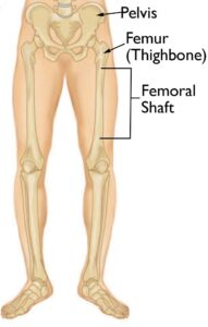
The femur, or thighbone, is the longest and most robust bone in the human body, making it highly resistant to fractures. However, significant force, such as that experienced in car accidents, is often required to break this bone, making such incidents the leading cause of femur fractures. The femoral shaft refers to the long, straight portion of the thighbone, and any break along this section is known as a femoral shaft fracture. This type of fracture generally necessitates surgical intervention for proper healing and recovery.
The femoral shaft runs from below the hip to where the bone begins to widen at the knee.
Types of Femoral Shaft Fractures
Femoral shaft fractures can vary significantly based on the force that led to the break. Bone fragments may remain aligned, known as a stable fracture, or may become misaligned, referred to as a displaced fracture. The injury may also be categorized by whether the surrounding skin remains intact (closed fracture) or if the bone penetrates through the skin (open fracture).
To categorize these fractures, doctors use specific classification systems based on factors such as:
- Fracture location: The femoral shaft is divided into three parts – distal, middle, and proximal.
- Fracture pattern: The break can occur in various directions, including horizontal, vertical, or angled.
- Injury severity: This includes whether the injury tears through the muscle and skin above the bone.
The main types of femoral shaft fractures include:
- Transverse fracture: Characterized by a horizontal break across the femoral shaft.
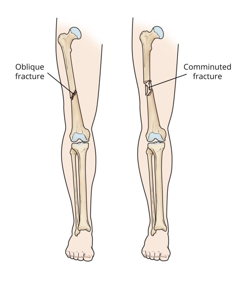
- Oblique fracture: Defined by an angled line running through the shaft.
- Spiral fracture: The break wraps around the shaft in a spiral pattern, often caused by a twisting force.
- Comminuted fracture: The bone is broken into three or more fragments.
- Open fracture: When bone fragments break through the skin or are exposed due to an external wound. Open fractures can cause extensive damage to muscles, tendons, and ligaments and carry a higher risk of complications, including infections, which may slow the healing process.
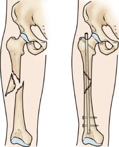
(Left) An oblique fracture has an angled line across the shaft. (Right) A comminuted fracture is broken into three or more pieces.
Causes
In younger individuals, femoral shaft fractures typically result from high-impact accidents. The primary causes include motor vehicle or motorcycle collisions, being struck by a vehicle while walking, falls from significant heights, and gunshot injuries. For older adults with weakened bones due to conditions like osteoporosis, even a low-impact fall from standing height can lead to a femoral shaft fracture.
Symptoms
A femoral shaft fracture generally produces intense, immediate pain. The affected leg will be unable to bear weight, and it may appear visibly deformed, possibly shorter or misaligned compared to the uninjured leg.
Doctor Examination
A thorough examination is essential for diagnosing and understanding a femoral shaft fracture.
Medical History and Physical Examination
To properly evaluate your injury, your doctor needs detailed information on how the injury occurred. For example, if it happened in a car accident, knowing specifics such as your speed, whether you were driving or a passenger, seat belt usage, and airbag deployment helps determine the mechanics of the injury and assess if other injuries may be present.
Additionally, your doctor will want to know about any pre-existing health conditions, including allergies, asthma, diabetes, or high blood pressure, and will ask about your use of tobacco products and any medications.
Following a discussion of your medical history, your doctor will conduct a thorough physical exam, focusing on your leg. They will look for:
- Visible deformities in the thigh or leg, such as unusual angles, twisting, or shortening.
- Open wounds or breaks in the skin.
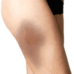
- Bruising or bony fragments pressing against the skin.
Your doctor will also palpate along the thigh, leg, and foot to:
- Identify any irregularities.
- Assess skin and muscle tightness around the thigh.
- Check for pulses.
- Test sensation and movement in the leg and foot (if you are conscious and alert).
Imaging Tests
Imaging tests provide critical details about the injury.
- X-rays: The primary method for assessing fractures, X-rays offer clear images of dense structures, such as bones. They reveal whether the bone is intact or broken, the fracture type, and its exact location within the femur.
- Computerized Tomography (CT) Scans: If X-rays do not provide enough information, a CT scan may be ordered. CT scans create cross-sectional images of the limb, offering a detailed view that can reveal the severity of the fracture and detect subtle fractures that may not appear clearly on X-rays, such as thin fracture lines.
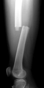
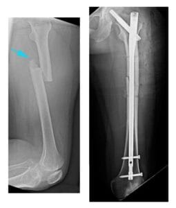
X-ray shows a transverse fracture of the femur. The break is a straight horizontal line across the shaft.
Treatment Options
Nonsurgical Treatment
In most cases, surgery is required to treat femoral shaft fractures effectively. It is rare for these fractures to heal without surgical intervention. Young children may sometimes be treated with a body cast; for more information, refer to resources on femur fractures in children.
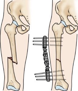
An external fixator is a temporary treatment often used when a patient has multiple injuries and is not ready for a longer surgical procedure. This device, which provides immediate stability to the fractured femur, consists of metal rods and pins that secure the bones in place from outside the body. Although an external fixator can remain on until the bone heals, it is generally used only temporarily until the patient is ready for definitive surgery.
Intramedullary Nailing
Intramedullary nailing is currently the preferred method for treating femoral shaft fractures. This procedure involves inserting a specially designed metal rod, or nail, into the femur’s canal. The nail spans the fracture site, stabilizing the bone and promoting proper alignment during healing.
Surgical Treatment
Timing of Surgery
Femoral shaft fractures are typically treated within 24 to 48 hours. However, surgery may be delayed if other life-threatening injuries or unstable medical conditions require immediate attention. Open fractures are treated with antibiotics upon hospital arrival to lower the infection risk, and the wound, surrounding tissue, and bone are thoroughly cleaned during surgery.
Between the initial emergency care and surgery, the doctor may place the injured leg in a long-leg splint or traction to maintain bone alignment and leg length. Skeletal traction involves a system of weights and counterweights that keep the leg straight and help relieve pain by holding the fractured bones together.
External Fixation
In some cases, metal pins or screws are surgically inserted into the bone above and below the fracture. These pins and screws connect to a stabilizing bar outside the skin, which holds the bones in proper alignment until the fracture can be permanently fixed.
External fixation is often used to hold the bones together temporarily when the skin and muscles have been injured.
Intramedullary Nail Placement
The intramedullary nail can be inserted at either the hip or knee end of the femur. Screws positioned above and below the fracture maintain proper alignment as the bone heals. Intramedullary nails are typically made of titanium and come in various sizes to fit different femur dimensions.
Intramedullary nailing provides strong, stable, full-length fixation.
Plates and Screws
In some situations, such as when a fracture extends into the hip or knee joint, intramedullary nailing may not be an option. Instead, the fractured bone fragments are realigned (reduced) and secured using screws and metal plates placed along the outer bone surface. This method effectively stabilizes complex fractures, particularly near joint areas.
(Left) This X-ray, taken from the side, shows a transverse fracture of the femur. (Right) In this front view X-ray, the fracture has been treated with intramedullary nailing.
Recovery Timeline
Femoral shaft fractures typically require 3 to 6 months to fully heal, though healing may take longer in cases of complex fractures, such as those that are open or involve multiple bone fragments, or in patients who smoke.
Pain Management
Pain is a natural aspect of the recovery process after an injury or surgery. Your healthcare team will focus on minimizing pain to help accelerate healing. Medications for short-term pain relief are commonly prescribed after surgery, and options include:
- Acetaminophen
- Nonsteroidal anti-inflammatory drugs (NSAIDs)
- Gabapentinoids
- Muscle relaxants
- Opioids
- Topical pain medications
Your doctor may combine these medications to enhance pain control while minimizing opioid use. Be aware that some pain medications may cause side effects, potentially affecting activities like driving. Your doctor will review possible side effects with you.
Use Caution With Opioids
While opioids can provide effective pain relief after surgery or injury, they carry a risk of dependency and overdose. Due to the risk of addiction, it’s crucial to use opioids only as directed and discontinue them as soon as your pain improves. If you find your pain does not lessen after a few days, consult your doctor for guidance.
Weight-Bearing Guidelines
Many doctors advocate for early movement of the leg during recovery. It is essential to follow your doctor’s instructions on weight-bearing to ensure proper healing. In most cases, patients are encouraged to bear as much weight as possible immediately after surgery, though full weight-bearing may need to wait until healing progresses. Initially, you will likely use crutches or a walker for additional support.
Physical Therapy
Since the injury may lead to muscle weakness, exercises are crucial for regaining strength, flexibility, and normal range of motion. Physical therapy is a key component of rehabilitation, helping to restore muscle function and manage post-surgical pain. Your physical therapist will teach you specific exercises during your hospital stay and will guide you in using assistive devices like crutches or a walker.
Potential Complications
Complications from Femoral Shaft Fractures
Femoral shaft fractures can lead to additional injuries or complications. Although rare, sharp bone fragments can damage surrounding nerves or blood vessels. One serious complication is acute compartment syndrome, a condition where pressure builds within the muscles, reducing blood flow and oxygenation to muscle and nerve cells. This requires emergency surgery, where incisions are made in the skin and muscle coverings to release the pressure.
Open fractures expose the bone to the external environment, increasing the risk of infection, which may necessitate multiple surgeries and long-term antibiotic treatment. Additionally, knee ligaments may sustain injuries during a femoral shaft fracture. Inform your doctor if you experience knee pain after surgery.
Complications from Surgery
Beyond typical surgical risks, such as blood loss or anesthesia-related complications, surgical treatment of femoral shaft fractures carries additional risks, including:
- Infection
- Nerve or blood vessel damage
- Blood clots
- Fat embolism (where bone marrow enters the bloodstream and may travel to the lungs, sometimes even occurring from the fracture alone)
- Malalignment, or improper alignment of bone fragments
- Delayed or failed healing (delayed union or nonunion)
- Hardware irritation, where the metal nails or screws used in repair may cause discomfort in the surrounding muscles or tendons

