Understanding ACL Injuries: Causes, Risks, and Treatment Options

Anterior cruciate ligament (ACL) injuries are among the most frequent knee injuries, often involving a sprain or tear. These injuries are particularly common among athletes engaged in high-intensity sports such as soccer, basketball, and football, where sudden stops and directional changes put the knee at risk. If you have sustained an ACL injury, surgical intervention may be necessary to restore full knee functionality. However, the need for surgery depends on various factors, including the extent of the injury and your activity level. For a comprehensive discussion on treatment options, refer to the article, “ACL Injury: Does It Require Surgery?” which serves as a valuable companion to this guide.

Anatomy of the Knee Joint

The knee joint is a complex structure where three bones converge: the femur (thighbone), tibia (shinbone), and patella (kneecap). Positioned at the front of the joint, the kneecap offers essential protection to the knee.

Ligaments play a crucial role in connecting bones and ensuring joint stability. In the knee, four primary ligaments function like robust ropes, holding the bones together and maintaining the joint’s stability.

Collateral Ligaments: Supporting Side-to-Side Movement

The collateral ligaments are located on either side of the knee. The medial collateral ligament (MCL), positioned on the inner side, and the lateral collateral ligament (LCL), located on the outer side, manage the knee’s side-to-side motion. These ligaments also provide support against excessive sideways movements, ensuring the knee remains stable.

Cruciate Ligaments: Controlling Forward and Backward Motion

Situated within the knee joint, the cruciate ligaments form an X-shape by crossing over each other. The anterior cruciate ligament (ACL) is positioned at the front, while the posterior cruciate ligament (PCL) is at the back. Together, they regulate the forward and backward movement of the knee.

The ACL runs diagonally through the center of the knee, preventing the tibia from sliding too far forward relative to the femur and ensuring rotational stability. The PCL, which is stronger and less frequently injured than the ACL, prevents excessive backward movement of the tibia, safeguarding the knee’s structural integrity.
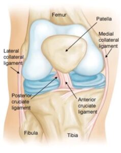
 

Normal knee anatomy.  The knee is made up of four main things: bones, cartilage, ligaments, and tendons.

ACL Injury Overview: Understanding Damage Severity

Approximately half of all anterior cruciate ligament (ACL) injuries occur in conjunction with damage to other knee structures, such as the articular cartilage, meniscus, or additional ligaments. Ligament injuries, classified as sprains, are assessed using a severity grading system to guide diagnosis and treatment.

Grade 1 Sprains: Mild Damage

In a Grade 1 sprain, the ligament sustains minor damage, resulting in slight stretching. Despite this, the ligament retains its ability to stabilize the knee joint effectively.

Grade 2 Sprains: Partial Tears

Grade 2 sprains occur when the ligament is stretched to the point of looseness, commonly referred to as a partial tear. This level of injury compromises the ligament’s stability function.

Grade 3 Sprains: Complete Tears

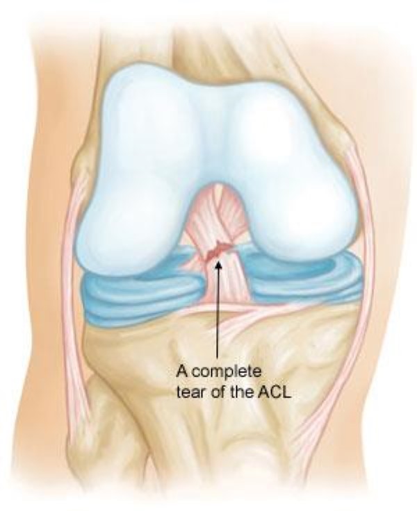
A Grade 3 sprain represents the most severe form of ligament injury, often described as a complete tear. In this case, the ligament is either torn into two pieces or detached entirely from the bone, leading to significant knee joint instability.

Partial vs. Complete ACL Tears

Partial tears of the ACL are relatively uncommon. The majority of ACL injuries involve complete or nearly complete tears, emphasizing the importance of prompt diagnosis and appropriate treatment to restore knee stability.


Injured ligaments are considered sprains and are graded on a severity scale.


Causes of ACL Injuries: Key Risk Factors

The anterior cruciate ligament (ACL) can be injured through various activities and situations, including:

  • Rapidly changing direction
  • Abruptly stopping movement
  • Decelerating while running
  • Improper landing technique after a jump
  • Direct impact or collision, such as during a football tackle

Research indicates that female athletes are at a higher risk of ACL injuries compared to male athletes in certain sports. This increased risk is attributed to factors such as differences in physical conditioning, muscle strength, and neuromuscular control. Other contributing elements may include variations in pelvis and lower limb alignment, greater ligament laxity, and the influence of estrogen on ligament structure and properties.

Symptoms of an ACL Injury: What to Watch For

When an ACL injury occurs, you may experience a combination of symptoms that signal the severity of the damage:

  • Audible “Pop” and Knee Instability: Many individuals report hearing a popping sound at the time of injury, often accompanied by a sensation of the knee giving way.
  • Pain and Swelling: Swelling usually develops within 24 hours. While the pain and swelling might subside over time if untreated, returning to physical activity with an unstable knee can lead to further damage, particularly to the meniscus.
  • Reduced Range of Motion: Injured individuals may struggle to fully extend or bend their knee.
  • Joint Line Tenderness: The knee may feel sensitive or sore along the joint line.
  • Walking Discomfort: Moving around or bearing weight on the injured leg can cause significant discomfort.
  • Doctor Examination: Assessing ACL Injuries

    Physical Examination and Patient History

During your initial consultation, the doctor will focus on understanding your symptoms and medical history to evaluate the severity of your knee injury. A thorough physical examination of the injured knee will be conducted, with comparisons made to the non-injured knee. In most cases, a detailed physical assessment is sufficient to diagnose ligament injuries, including an ACL tear.

Imaging Tests for Confirmation

To support the diagnosis, your doctor may recommend imaging tests, including:

  • X-rays: While X-rays do not reveal soft tissue injuries such as an ACL tear, they can help identify fractures or bone injuries associated with the trauma.
  • Magnetic Resonance Imaging (MRI): An MRI provides detailed images of soft tissues, including the ACL, meniscus, and cartilage. Although not always necessary for diagnosing a torn ACL, an MRI can help identify additional injuries to other soft tissue structures in the knee.

Treatment Options for ACL Tears

Treatment strategies for an ACL tear are tailored to the patient’s unique needs, including age, activity level, and overall health.

  • Surgical Intervention: Active individuals, such as young athletes engaged in agility-based sports, typically require surgery to safely resume their activities.
  • Non-Surgical Management: Older or less active individuals may manage their condition effectively without surgery, returning to a less strenuous lifestyle with the help of physical therapy and other conservative treatments.

For more detailed insights, continue to the related guide, “ACL Injury: Does It Require Surgery?”

Guidelines for Managing ACL Injuries

To assist healthcare professionals, the American Academy of Orthopaedic Surgeons (AAOS) has developed evidence-based guidelines to optimize the management of ACL injuries.

Scroll to Top