Understanding the Condition
Who Gets These Fractures?
- Younger Patients: Typically, from high-energy injuries like car accidents or falls from significant heights.
- Older Patients: This is often due to weaker bones (osteopenia), which makes them more susceptible to fractures even from minor falls.
Common Features
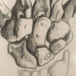
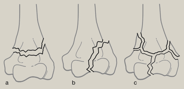
- Comminution: This means the bone is broken into several pieces, making the fracture complex.
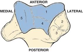
- Complex Bone Structure: The intricate shape of the distal humerus (the lower part of the upper arm bone) adds to the challenge of treating these fractures.
Initial Evaluation Goals
- Identify the Fracture Pattern: Understand how the bone is broken.
- Check for Previous Elbow Problems: Look for any existing elbow issues that might affect treatment.
- Assess Soft Tissue Damage: Determine if open wounds or significant soft tissue injuries exist.
- Identify Other Injuries: Look for related injuries in the arm or nearby nerves and blood vessels.
Diagnostic Imaging
- X-rays: First step to visualise the fracture. Images are taken from the front (anteroposterior) and side (lateral) views.
- X-rays help identify fracture lines, fragments, and associated injuries in nearby bones.
- Due to the complexity of the distal humerus, X-rays might need to show the full extent of the fracture clearly.
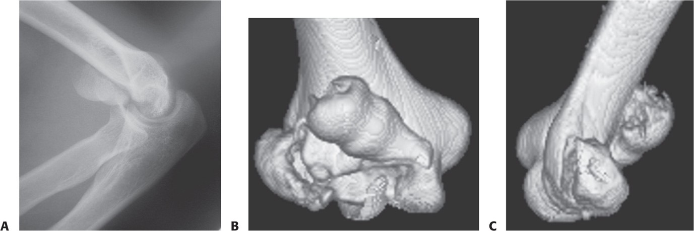
- CT Scans with 3D Reconstruction: Provides a detailed view of the fracture, helping the surgeon plan the surgery more accurately.
- Shows the exact configuration of broken bone pieces and their positions
.
- Traction Radiographs: Taken in the operating room under anaesthesia to get a clearer picture if CT scans are unavailable.
Surgical Treatment
Internal Fixation
- Preferred Treatment: Most distal humerus fractures are treated with internal fixation.
- Modern Techniques: Include strategies to improve stability using pre-shaped plates and screws that lock into the plates.
- Elbow Replacement: This is considered for elderly patients with pre-existing elbow problems or very complex fractures due to weak bones.
- Goal: Achieve a stable bone construct that allows immediate movement without the risk of the fracture moving out of place again.
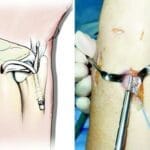
Fixation Techniques
Use of Plates and Screws:
- Plates are applied on both sides of the humerus to maximise fixation.
- Screws lock into these plates to provide additional stability.
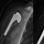
Surgical Example:
- Parallel Plate Fixation: Two plates are placed on the humerus’s medial (inner) and lateral (outer) sides.
- Post-surgery X-ray: This shows the bone pieces perfectly aligned and securely fixed using plates, with a clear picture of any additional repairs, like fixing an olecranon osteotomy (a cut made in the elbow bone to allow better access during surgery).
Summary
Understanding the fracture type, the patient’s history, and the extent of injuries are crucial steps before surgery. Advanced imaging techniques like CT scans help in accurately planning the surgery. The primary goal of surgery is to stabilise the bone using plates and screws, enabling immediate movement and reducing the risk of complications. For elderly patients with complex fractures, elbow replacement may be considered as an alternative. Proper surgical techniques ensure that even the most complex fractures can be treated effectively, allowing for a smooth recovery.