Introduction
A bunionette, also known as a tailor’s bunion, is a bony prominence that forms on the outer side of the foot, similar to a bunion but affecting the fifth toe. The term “tailor’s bunion” originates from the historical practice of tailors sitting cross-legged for extended periods, causing repeated pressure and friction along the outer edge of their feet. This constant pressure led to the formation of calluses and irritation at the base of the fifth toe.
This guide provides essential insights into:
- The location and development of a bunionette
- The issues it can cause
- Available treatment options
Anatomy
Where Does a Bunionette Develop?
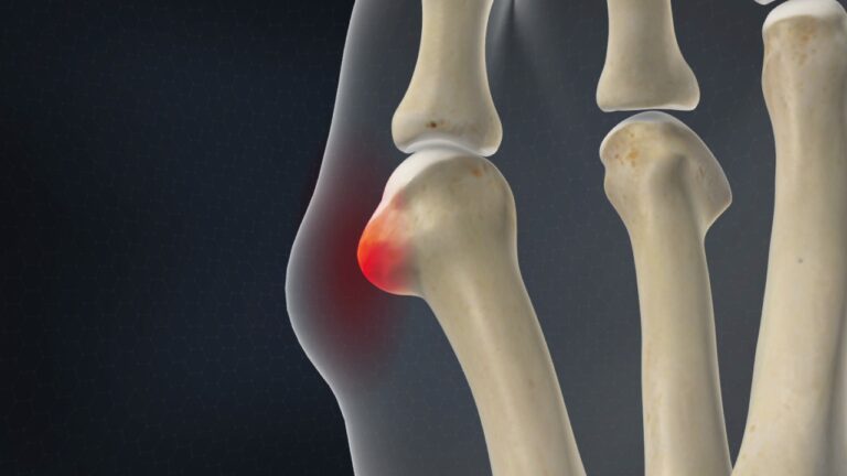
A bunionette forms at the metatarsophalangeal (MTP) joint, the area where the fifth toe connects to the foot. This joint comprises the metatarsals, the long bones in the foot, and the phalanges, the small bones that make up the toes. While the big toe has two phalanges, each of the other toes, including the fifth toe, has three phalanges.
Causes
How Does a Bunionette Develop?
A bunionette typically forms due to excessive pressure on the fifth metatarsal head, often caused by wearing shoes that are too narrow. Over time, this pressure leads to the formation of an abnormal bony prominence that rubs against footwear, resulting in irritation, callus formation, and tissue thickening.
As people age, their feet may widen, causing them to splay outward. If narrow footwear is continued, this can lead to the development of both a bunion on the inner side of the foot and a **bunionbunionette on the outer side. T
Any bony protrusion furtherReducing pressure is the key tosurgical removal of the bony
Symptoms
What Do Bunionettes Feel Like?
A bunionette commonly causes pain and discomfort, particularly when wearing shoes that press against the affected area. Many individuals experience difficulty finding footwear that does not exacerbate the pain. Additionally, the visible swelling and prominence of the bunionette can be considered aesthetically undesirable by some.
Diagnosis
How Do Doctors Identify a Bunionette?
A bunionette is typically diagnosed through a physical examination, as its presence is often visually apparent. X-rays may be used to assess whether the foot has widened (splayed) and to determine if surgical intervention may be necessary. These imaging studies help evaluate the severity of the deformity and guide treatment planning.
Treatment
What Can Be Done for a Bunionette?
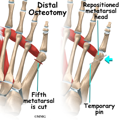
If the angle of the fifth metatarsal is significantly increased, your doctor may recommend osteotomy, a surgical procedure in which the metatarsal bone is cut and realigned. Once the osteotomy is performed, the bones are stabilized using metal pins, which remain in place during the healing process.
Nonsurgical Treatment
The first line of treatment focuses on proper footwear that accommodates the width of the forefoot, reducing pressure on the bunionette. Additional conservative measures include:
- Protective pads: Specially designed doughnut-shaped pads can help redistribute pressure and alleviate discomfort. These are commonly available in drugstores and supermarkets.
Surgical Treatment
If conservative treatments fail to provide relief, surgery may be considered to correct the deformity and relieve pressure. Surgical options include:
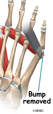
Bunionette Removal
This procedure involves:
- Making a small incision over the bunionette.
- Removing the bony prominence using a chisel.
- Smoothing the bone edges to minimize irritation.
- Closing the incision with small stitches.
In cases where the foot has splayed, surgery may also involve realigning the fifth metatarsal to restore proper foot structure.
Rehabilitation
What Should I Expect After Treatment?
Nonsurgical Rehabilitation
For patients with a painful bunionette, four to six physical therapy sessions may provide relief. A physical therapist can recommend footwear with a wider toe box, ensuring the metatarsals are not compressed. Additionally, a protective pad can be placed over the bunionette to reduce pressure and discomfort.
With these footwear adjustments, normal walking can often resume immediately. However, high-impact activities should be minimized for several weeks to allow inflammation to subside.
Physical therapy treatments may include:
- Ultrasound therapy, moist heat, and soft-tissue massage to manage pain and swelling.
- Iontophoresis, a technique using mild elect
Post-Surgical Recovery
After surgery, patients are typically fitted with a post-op shoe featstiff, wooden sole that prevents excessive foot movement, protecting the healing site. Metal pins, if used, are usually removed after three to four weeks, once the bone has begun to mend.
Recovery Timeline
- Crutches may be necessary for a short period, with physical therapy guidance on proper use.
- A bandage or dressing is typically worn for about a week.
- Stitches are removed 10 to 14 days post-surgery unless absorbable sutures were used.
- Follow-up X-rays help monitor bone healing and assess the correction achieved.