Understanding Patellar Fractures: Causes, Symptoms, and Treatment Options
Patellar fractures can present in a variety of patterns. The break may be a clean, straightforward split into two pieces or a more severe injury where the kneecap shatters into multiple fragments.
- Direct falls onto the knee
- High-impact trauma, such as a football tackle where a helmet strikes the kneecap or a high-speed impact from a baseball or softball
- Collision injuries, such as hitting the dashboard with the kneecap during a car accident
- Non-Surgical: Simple fractures, where the bone remains in alignment, can often be managed with a cast or splint, allowing the bone to heal naturally over time.
- Surgical: For more complex fractures, where the bone fragments are displaced, surgical intervention is typically required. This procedure realigns and stabilizes the kneecap, ensuring proper healing and the restoration of knee function.
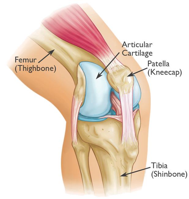
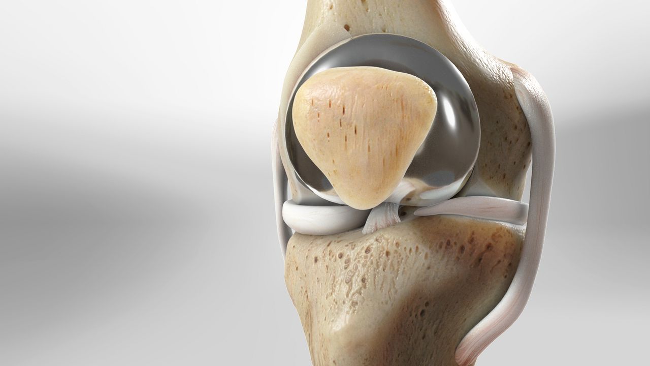
The patella covers and protects the knee joint.
Description of Patellar Fractures
Patellar fractures can present in a variety of patterns. The break may be a clean, straightforward split into two pieces or a more severe injury where the kneecap shatters into multiple fragments. These fractures can occur at different locations on the patella, such as the upper, middle, or lower sections. In some cases, multiple fractures can occur simultaneously, affecting more than one area of the kneecap. Understanding the type and location of a patellar fracture is essential for determining the most effective treatment approach.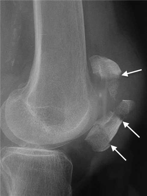
This X-ray of a knee taken from the side shows a patella that has been fractured in three places.
Stable Fracture
A stable patellar fracture is a type of nondisplaced fracture, meaning the broken bone pieces remain largely aligned. The fragments may either stay in contact or have a minimal separation of just one or two millimeters. In this type of fracture, the stability of the bone ensures that it remains in proper alignment throughout the healing process, often requiring less invasive treatment methods. Stable fractures typically respond well to conservative management, such as immobilization with a cast or splint, allowing the natural healing process to restore the bone's integrity.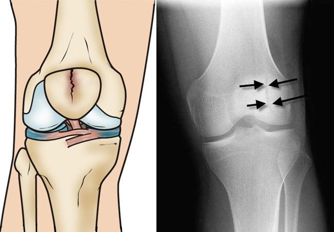 Illustration and X-ray show a vertical, stable fracture of the patella.
Displaced Fracture
A displaced patellar fracture occurs when the broken ends of the bone are misaligned and separated. This misalignment disrupts the normally smooth surface of the knee joint, potentially impairing its functionality and causing pain during movement. Due to the severity of this fracture type, surgical intervention is often required to realign the bone fragments and restore the joint's normal structure. This ensures proper healing and helps prevent long-term complications, such as joint stiffness or arthritis.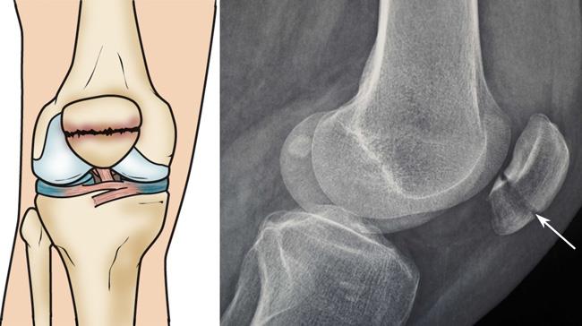 Illustration and X-ray show a front (left) and side (right) view of a two-part fracture across the patella (transverse fracture) with slight displacement between the broken pieces of bone.
Comminuted Fracture
A comminuted fracture occurs when the patella breaks into three or more fragments. The stability of this type of fracture depends on its specific pattern. It may be classified as either stable, where the bone fragments remain aligned, or unstable, where the pieces are misaligned and require surgical intervention.Open Fracture
An open fracture is a severe injury where the broken bone protrudes through the skin or a wound penetrates down to the bone. This type of fracture often causes extensive damage to the surrounding soft tissues and requires a longer healing time due to its complexity. Open fractures are particularly dangerous because the broken skin increases the risk of infection in both the wound and the exposed bone. Immediate medical attention is crucial to clean the wound, prevent infection, and initiate appropriate treatment to promote healing and minimize complications.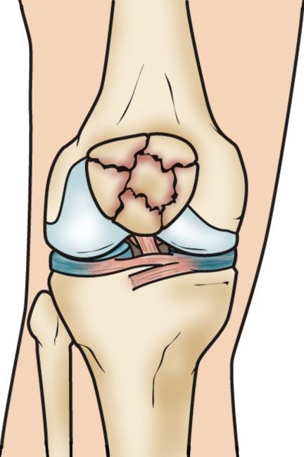 A comminuted fracture
Causes of Patellar Fractures
Patellar fractures typically result from direct or indirect trauma to the knee. Common causes include: Direct Impact: Falling directly onto the knee or sustaining a sharp blow, such as in a head-on car collision where the kneecap strikes the dashboard. Indirect Trauma: Sudden, forceful contraction of the quadriceps muscle can indirectly fracture the patella by pulling it apart.Symptoms of Patellar Fractures
The most recognizable symptoms of a patellar fracture include: Pain and Swelling: Localized in the front of the knee. Bruising: May occur around the knee joint. Movement Impairment: Inability to straighten the knee, perform a straight leg raise, or maintain knee extension. Weight-Bearing Issues: Difficulty or inability to stand or walk.Doctor Examination and Diagnosis
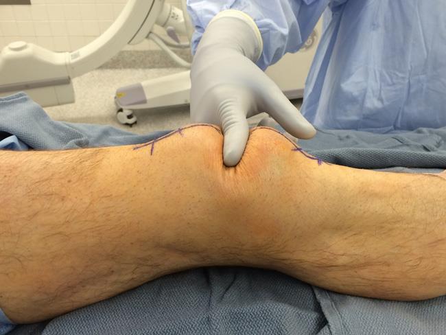
Physical Examination
Your doctor will begin by reviewing your symptoms and medical history, followed by a thorough examination of the knee. Key aspects include: Fracture Detection: The edges of the fracture are often palpable through the skin, especially in displaced fractures. Checking for Hemarthrosis: This condition involves the collection of blood in the joint space due to the fracture, leading to significant swelling. If needed, your doctor may perform a procedure to drain the blood and alleviate pain.Imaging Tests
To confirm the diagnosis and assess the fracture's severity, X-rays are typically ordered. These images provide detailed information about the fracture pattern and help guide the treatment plan. Early diagnosis and treatment are critical to managing patellar fractures effectively and ensuring optimal recovery.The significant displacement of this patient's fracture has created a large gap between the pieces of bone.
X-Rays: An Essential Tool for Diagnosing Patellar Fractures
X-rays are a vital imaging technique used to visualize dense structures like bones. When diagnosing a patellar fracture, your doctor will order X-rays from multiple angles to assess the break and evaluate the alignment of the bone fragments.Identifying Bipartite Patella
In rare cases, a person may have a congenital condition known as bipartite patella, where the patella consists of two or more separate bone pieces that did not fuse together during development. This condition can sometimes be mistaken for a fracture on an X-ray. To differentiate between a fracture and a bipartite patella, your doctor may: Examine the X-ray images for characteristic signs of bipartite patella. Take an X-ray of the opposite knee, as many individuals with bipartite patella have the condition in both knees. By carefully analyzing the X-rays, your doctor can ensure an accurate diagnosis and tailor the treatment approach accordingly.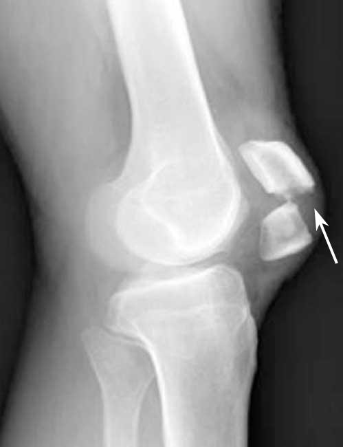 This X-ray of a patellar fracture shows significant displacement between the broken pieces of bone.
Nonsurgical Treatment
In cases where the broken bone fragments are not displaced, surgery may not be necessary. Instead, your doctor may use a cast or splint to immobilize the knee, keeping it straight to prevent movement. This approach ensures that the bone fragments remain properly aligned during the healing process. Weight-Bearing Restrictions: Depending on the type of fracture, your doctor may allow partial weight-bearing on the affected leg while wearing the cast or brace. However, with certain fractures, weight-bearing may be prohibited for 6 to 8 weeks. Your doctor will provide detailed instructions based on your condition.Surgical Treatment
If the bone fragments are displaced, surgical intervention is usually required to realign and stabilize the patella. Displaced fractures often struggle to heal naturally, as the strong quadriceps muscles attached to the patella can pull the fragments apart during the healing process.Timing of Surgery
Closed Fractures: If the skin remains intact, surgery may be delayed until any abrasions or scrapes around the knee have healed. Open Fractures: If the fracture pierces the skin, immediate surgery is necessary to minimize the risk of infection. The wound and bone are thoroughly cleaned, and the fracture is typically repaired during the same surgical procedure. Surgical Procedures The type of surgery performed depends on the nature and severity of the fracture.Transverse Fracture Repair
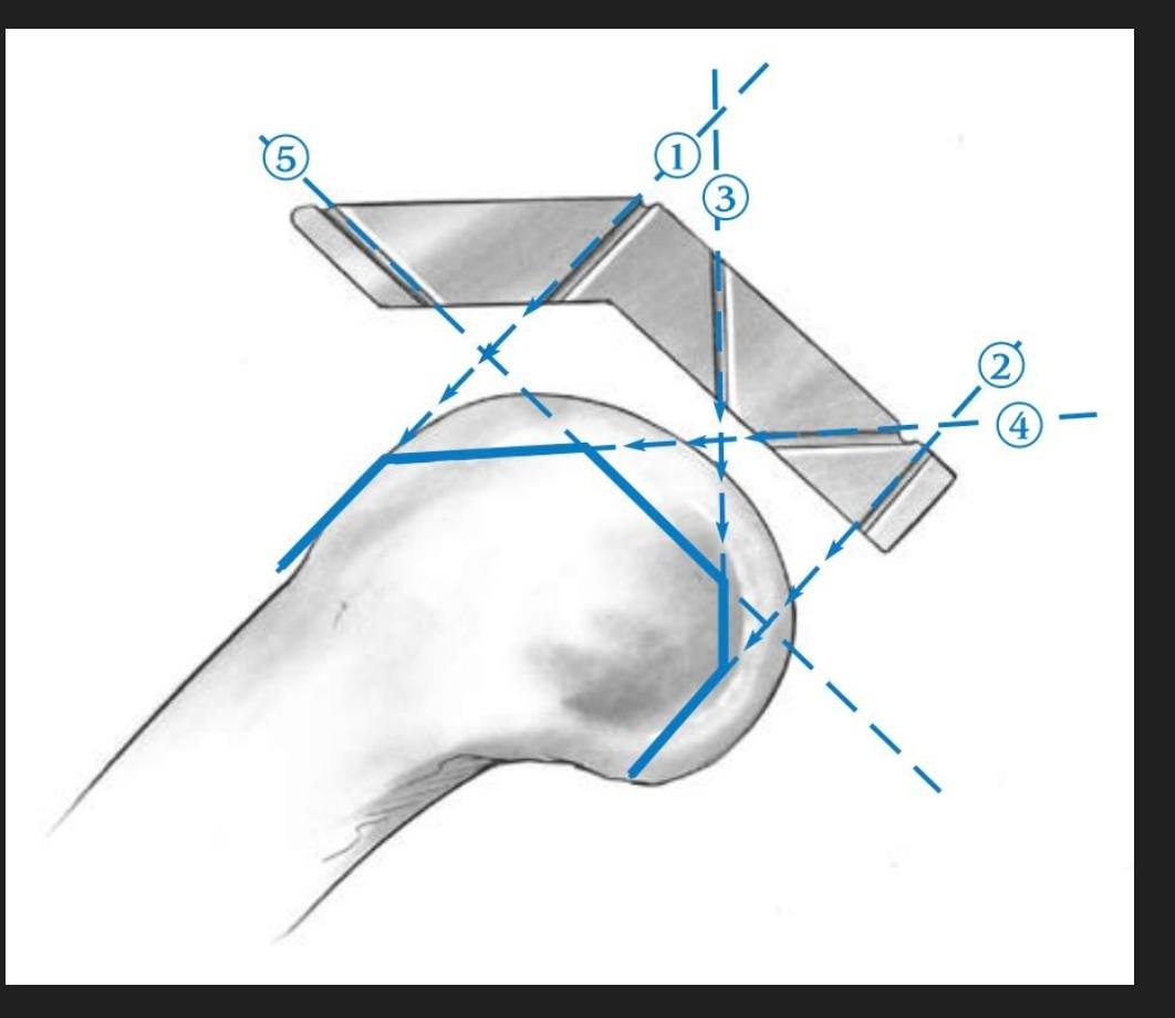
For fractures that split the patella into two parts, surgeons often use screws or a combination of pins and wires arranged in a figure-of-eight tension band. This technique compresses the two fragments together, promoting healing. This method is most effective for fractures near the center of the patella but is less suitable for fractures at the ends or comminuted fractures (those with multiple pieces), as over-compression can occur.Alternative Fixation Methods
Small screws or a combination of screws and plates may be used as an alternative, particularly for fractures that cannot be stabilized effectively with a tension band. Your doctor will discuss the most appropriate surgical approach for your specific fracture, along with any potential risks or complications associated with the procedure. Early and effective treatment is crucial for restoring knee function and minimizing long-term issues.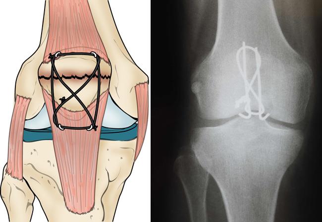 In this illustration and X-ray, a figure-of-eight tension band has been used to hold a transverse fracture together.