TraumaPatient education
Understanding Osteoporosis and the Impact of Spinal Compression Fractures
Although vertebral compression fractures can have various causes, osteoporosis often plays a crucial role. For many individuals, experiencing such a fracture may be the first indicator of a weakened skeletal structure due to osteoporosis.
Published
2 November 2024
Reading time
6 min
Words
1,292
Sections
8

As we age, our bones naturally become thinner, and their strength gradually diminishes. Osteoporosis is a condition where bones weaken significantly, making them prone to fractures. This condition can progress silently over many years, often without noticeable symptoms until a fracture occurs.
In cases of osteoporosis, fractures commonly happen in the spine. Known as vertebral compression fractures, these spinal injuries affect approximately 1.5 million individuals annually in the United States, nearly twice as frequently as other osteoporosis-related fractures, like those of the hip or wrist.
Although vertebral compression fractures can have various causes, osteoporosis often plays a crucial role. For many individuals, experiencing such a fracture may be the first indicator of a weakened skeletal structure due to osteoporosis.

Anatomy
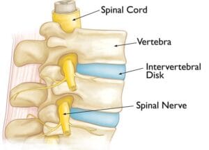
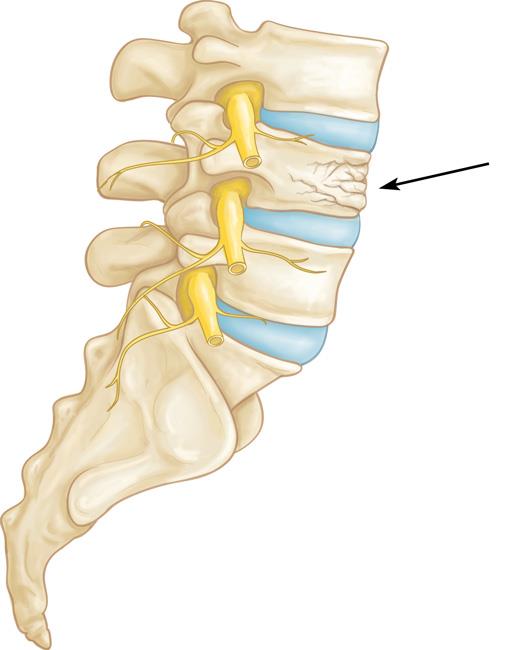
The spine consists of 24 stacked bones, known as vertebrae, which together form a protective canal for the spinal cord. In addition to the vertebrae, the spine includes:- Spinal Cord and Nerves: These "electrical cables" run through the spinal canal, transmitting signals between the brain and muscles. Nerve roots extend outward from the spinal cord through openings in the vertebrae.
- Intervertebral Discs: Positioned between the vertebrae, these flexible, flat discs'each about half an inch thick'act as cushions, absorbing shock and protecting the vertebrae during activities like walking and running.
Causes
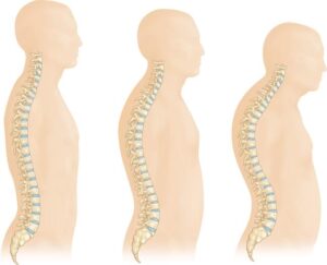
Osteoporosis is a natural part of aging, as bones gradually lose strength over time. When the vertebrae in the spine weaken, they may become thinner and flatter, often resulting in a loss of height and a rounded back or hunched posture in elderly individuals. These weakened vertebrae are particularly susceptible to fractures. A vertebral compression fracture occurs when excessive force is placed on a weakened vertebra, causing it to crack and reduce in height. While these fractures are commonly triggered by falls, people with osteoporosis can sustain them even from everyday movements like twisting, reaching, coughing, or sneezing.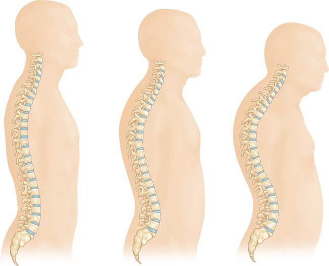
Symptoms Vertebral compression fractures often lead to localized back pain, usually centered around the fracture site. These fractures frequently occur around the waistline, as well as just above (mid-back) or below it (lower back). Movement, especially when changing positions, can intensify the pain, which typically eases with rest or lying down. Activities like coughing or sneezing may also increase discomfort. While it's less common, the pain can sometimes radiate to other areas, such as the abdomen or legs. In severe cases, nerve pain may extend into the legs if the fracture impacts nearby nerves.

Doctor Examination
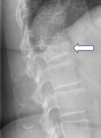
Physical Examination During your consultation, the doctor will review your general health, medical history, and specific symptoms. They'll conduct a physical exam to assess your spine's alignment and posture while standing, as well as press on specific areas of your back to determine if the pain originates from a muscle or bone injury. To rule out damage to spinal nerve roots, a neurological exam will assess for any loss of sensation, reflex changes, or muscle weakness. Imaging Tests Imaging tests are crucial for evaluating the fracture and determining whether it is recent (acute) or older (chronic).- X-rays: X-rays capture detailed images of dense structures like bones. If a vertebral compression fracture is suspected, X-rays can confirm the diagnosis. Additional spinal issues, such as degenerative disc disease (disc space narrowing) or scoliosis (sideways spinal curvature), may also be visible, especially in older adults.
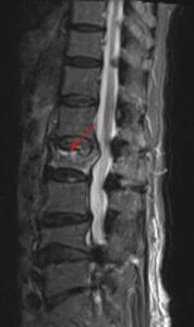
- Magnetic Resonance Imaging (MRI): An MRI provides a more detailed view of the surrounding soft tissues, including nerves and discs. MRIs are also valuable for estimating the fracture's age; a new fracture may appear brighter due to swelling or inflammation, while an older fracture will look more like the surrounding bone.
- Bone Scan: A bone scan detects unusual bone activity, including fractures, and can help differentiate between acute and chronic fractures.
- Computed Tomography (CT) Scan: A CT scan reveals both soft tissues and bone, enabling the doctor to determine if the fracture has spread into the spinal canal, where the spinal cord and nerve roots are located.
Bone Density Testing
When a vertebral compression fracture occurs, it's essential to evaluate whether osteoporosis is present and determine its severity. X-rays can often reveal overall bone thinning in the spine, a condition known as osteopenia, which precedes osteoporosis. Osteopenia indicates reduced bone mass, which, if untreated, can progress to osteoporosis, making bones much more fragile and prone to fractures. To measure the degree of bone loss, a Dual-Energy X-ray Absorptiometry (DEXA) scan is often used. This bone mineral density test helps the doctor assess fracture risk both in the spine and other bones. The DEXA results also guide treatment decisions, including whether medication is needed to address bone density loss. Timely osteoporosis treatment is critical; research shows that over 30% of individuals with a spinal compression fracture may experience another fracture within a year.Treatment
Nonsurgical Treatment
Many patients with vertebral compression fractures recover within three months without specific fracture repair. Basic care, including short periods of rest, limited use of pain relief medications, and sometimes wearing a brace to restrict movement, often suffices. It's important to note that while the bone heals, it doesn't regain its original shape, but the healed bone becomes stable and less likely to collapse further. If osteoporosis is diagnosed, the risk for additional vertebral fractures'as well as fractures in the hip or wrist'increases. During this period, your doctor will discuss treatment options to improve bone density and reduce fracture risk.Surgical Treatment
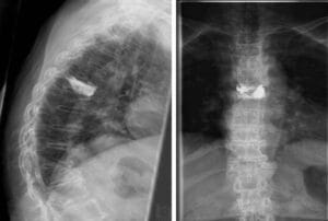
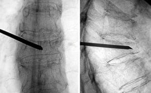
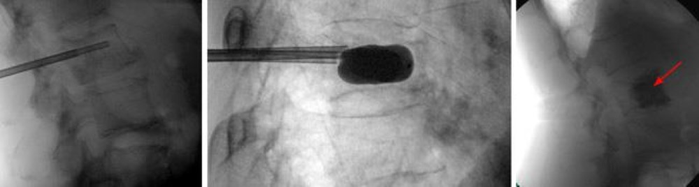
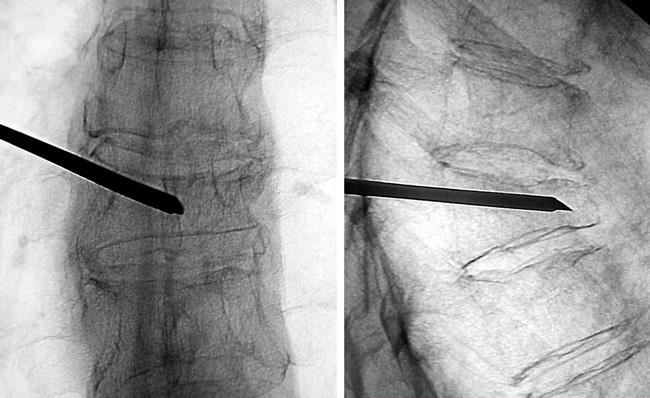
In cases where pain persists despite nonsurgical treatments, surgery may be an option. In the past, surgical solutions for vertebral compression fractures were extensive. However, minimally invasive vertebral augmentation techniques, such as kyphoplasty and vertebroplasty, are now available.- Kyphoplasty: This procedure involves inserting a needle into the fractured vertebra using X-ray guidance. Through the needle, a small balloon device, or balloon tamp, is placed in the vertebra and then inflated to restore the vertebra's height and shape. Once the balloon is removed, the cavity is filled with bone cement to reinforce the vertebra. Kyphoplasty can be done with general anesthesia or local anesthesia and typically allows patients to resume normal activities soon after surgery.

- Vertebroplasty: Similar to kyphoplasty, vertebroplasty involves injecting bone cement into the fractured vertebra. However, no balloon is used to create a cavity; instead, the cement is injected directly into the bone. This procedure is also done with the patient lying face down, enabling access to the vertebra from the back, and allows a quick return to regular activities.
Surgical Outcomes
Research on vertebral augmentation procedures suggests that the benefits of vertebroplasty may be short-lived, while kyphoplasty appears to improve function and alleviate pain more effectively and quickly than nonsurgical treatments. Your doctor will set realistic expectations for recovery and explain the anticipated outcomes if kyphoplasty or vertebroplasty is recommended.