Understanding Charcot Foot: A Diabetic Complication and Its Prevention

Diabetes, a chronic condition characterized by elevated blood sugar levels, impacts approximately 9% of the U.S. population—about 30 million individuals. Among its complications, diabetic foot issues pose a significant health challenge and are a leading cause of hospitalizations.

The majority of foot complications in diabetic individuals stem from the disease’s detrimental effects on small blood vessels. In the feet, this vascular damage results in two primary problems:

  1. Reduced Sensation: Impaired blood flow to the nerves diminishes sensation, making individuals less aware of cuts, injuries, or trauma to their feet.
  2. Delayed Healing: Poor circulation to the skin and underlying tissues hinders wound healing. This increases the risk of wounds becoming chronic, providing a gateway for bacterial infections, which may lead to severe diabetic foot infections.

While numerous treatment options exist for addressing diabetic foot complications, prevention remains the cornerstone of effective management. Maintaining strict blood sugar control and conducting daily foot inspections are essential strategies for reducing the risk of these debilitating conditions.

One severe consequence of diabetic neuropathy—a type of nerve damage caused by diabetes—is Charcot arthropathy (also referred to as Charcot neuroarthropathy or Charcot foot). This condition arises from unnoticed bone injuries in the foot, potentially resulting in significant deformities and mobility issues. The following sections will delve deeper into Charcot arthropathy, its implications, and management strategies.

Description

Charcot arthropathy, also known as Charcot foot, is a severe condition of the foot and ankle caused by an inability to sense injuries. This loss of sensation can result in progressive deformities. For Charcot foot to develop, neuropathy (nerve damage) must already be present, with diabetes being the most common underlying cause.

In individuals with diabetic neuropathy, fractures or dislocations in the foot may occur without the patient being aware. These injuries can result from:

  1. Traumatic Events: Obvious incidents such as falls or ankle sprains.
  2. Chronic Microtrauma: Gradual damage due to altered weight distribution on the foot. In individuals without neuropathy, such abnormal forces typically cause pain, prompting the person to adjust their posture and redistribute weight. However, diabetic patients may not feel pain or make these adjustments, allowing repeated stress to cause fractures and other injuries over time.

Without the ability to feel pain, a person with diabetes may continue walking on an injured foot, exacerbating the damage and leading to severe deformities. Interestingly, only 25–50% of individuals with Charcot foot recall a specific injury preceding the condition.
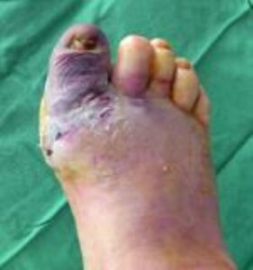
A severely infected big toe. This infection began two days earlier as a small blister at the tip of the toe.

Risk Factors

While neuropathy is a prerequisite for Charcot arthropathy, other factors can increase the likelihood of its development:

  1. Obesity: Excess body weight places additional strain on feet with impaired sensation, significantly raising the risk.
  2. Age: The risk of developing Charcot foot also increases as diabetic individuals grow older.

Impact of Deformities

The deformities caused by Charcot arthropathy can lead to several complications:

  • Difficulty Wearing Shoes: Severe deformities may make it impossible to find or wear normal footwear.
  • Loss of Stability: Deformities can destabilize the foot and ankle, hindering the ability to walk normally.
  • Increased Risk of Ulcers: The most critical concern is that deformities often result in areas where the bones protrude just beneath the skin. These pressure points can cause skin ulcers, significantly raising the risk of foot infections.

Diabetics face unique challenges in wound healing due to poor blood circulation and reduced immune response. In cases of Charcot foot, if an ulcer develops, the likelihood of amputation rises dramatically. Diabetics with Charcot foot have a 50% chance of requiring an amputation once an ulcer forms, compared to just a 10% risk without an ulcer.


(Left) This patient with Charcot of the ankle has developed a deformity that places abnormal pressure on the outside of the ankle. (Right) This pressure has led to the development of a chronic sore (ulcer) that can be extremely difficult to treat.


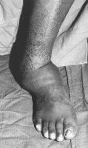
This patient with Charcot arthropathy has a severe deformity.


Symptoms

Charcot arthropathy often develops without significant pain, but patients may experience other noticeable symptoms.

  • Swelling: One of the earliest signs is unexplained swelling in the foot, often without any apparent injury. Many patients first notice difficulty fitting into their usual footwear.
  • Redness and Warmth: Inflammation, characterized by redness and warmth, is another early indicator. These changes occur as part of the body’s natural inflammatory response to underlying injuries in the foot.

These symptoms—swelling, redness, and warmth—can sometimes be mistaken for an infection. However, an infection is unlikely if the skin remains unbroken and no wounds are present. For a diabetic patient with a newly swollen foot and no wounds, Charcot arthropathy is the most probable diagnosis. A simple test to distinguish Charcot from an infection is to elevate the affected foot for several minutes. In Charcot, the redness, swelling, and warmth typically improve; in an infection, these symptoms usually persist.

Doctor Examination

Medical History and Physical Evaluation
During your visit, the doctor will inquire about your symptoms and overall health. Key questions may include:

  • Details about any possible foot injuries.
  • Information regarding your diabetes management, including your most recent hemoglobin A1C level and any history of foot wounds or infections.

Following the discussion, the doctor will conduct a thorough examination of your foot:

  • Inflammation Check: If your foot is red, swollen, and warm, the doctor may elevate it to see if the symptoms subside.
  • Inspection for Ulcers or Bony Prominences: They will examine the skin for any signs of ulcers or protruding bones that could cause pressure points leading to ulcers.
  • Foot Shape Analysis: The doctor will assess the foot’s shape for any visible deformities and determine if these are flexible (correctible) or rigid.
  • Blood Flow and Sensation Testing: Circulation and nerve function in the foot will also be evaluated.

Imaging Tests

  • X-rays: X-rays are a crucial diagnostic tool for Charcot foot. In the early stages, X-rays may appear normal. However, as the condition advances, signs such as fractures, bone fragments, and joint destruction become evident. Over time, more severe deformities, dislocations, and new bone formations may develop, indicating progressive Charcot arthropathy.
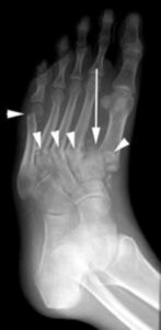
 

The patient shown in the X-ray had noticed swelling of the foot for approximately 3 weeks without any known injury. The X-ray shows several fractures (arrowheads) and a dislocation of the first metatarsal (arrow). Such a severe injury in patients without diabetes would be seen only after a high-energy trauma.

 

Computed Tomography (CT) Scan

A CT scan, often described as a three-dimensional X-ray, offers highly detailed images of the bones in the foot and ankle. This imaging technique is particularly useful for surgical planning in Charcot foot cases. If your doctor determines that surgery is necessary, a CT scan can help precisely map out the procedure, ensuring the most appropriate surgical approach is selected.

Magnetic Resonance Imaging (MRI) and Ultrasound

MRI and ultrasound imaging provide superior visualization of the soft tissues in the foot and ankle compared to X-rays. These tests are commonly ordered when an infection is suspected, especially in cases where distinguishing between inflammation from Charcot foot and a potential bone infection (osteomyelitis) is challenging. An MRI is particularly effective at identifying abscesses (pockets of infection) in soft tissues, which can be a critical factor in diagnosis and treatment.

Bone Scan and Indium Scan

Bone scans, a type of nuclear medicine imaging, are valuable tools for determining whether a bone infection is present. Depending on the specific issue, your doctor may choose from various types of bone scans. While these scans are slightly more effective than MRI at differentiating between Charcot foot and a bone infection, they can sometimes yield inconclusive results.

An indium scan, a specialized test, involves tagging white blood cells with a marker to track their movement. This technique helps identify whether the immune response is targeting the bone, indicating an infection. Both Charcot foot and bone infections can result in a positive bone scan, but only an infection will show a marked increase in activity on the indium scan, making it a critical tool for definitive diagnosis.

Treatment Goals

The primary objective in treating Charcot foot is to achieve an ulcer-free, stable, and plantigrade (flat-resting) foot that enables safe walking using commercially available diabetic footwear. The treatment approach focuses on the following goals:

  • Stability: Ensuring the foot and ankle have the structural strength to support body weight and facilitate walking without causing further deformities.
  • Plantigrade Position: The foot should rest flat on the floor, similar to a three-legged stool where the heel, big toe, and little toe evenly bear weight.
  • Braceability: Even if the foot’s position isn’t completely normal, it should be possible to make it stable and plantigrade using shoe inserts or braces to minimize deformities and ulcers.
  • Ulcer-Free Foot: Preventing ulcers is critical to avoiding infections and amputations. Addressing bony prominences under the skin helps prevent ulcers while promoting a functional walking ability.

Nonsurgical Treatment

  1. Casting:
    • In the early stages, casting or a special boot is used to protect the foot and ankle, allowing the bones to heal in a stable position and preventing further deformities.
    • A “total contact cast,” which covers the entire foot, is the most commonly used type for Charcot foot.
    • Patients must avoid putting weight on the foot until healing begins, often requiring crutches, a knee scooter, or a wheelchair. Healing can take up to three months or longer.
    • Casts are typically changed every one to two weeks to accommodate reduced swelling and to prevent ulcers.
    • Once swelling subsides, patients may transition to a removable, heavy-duty boot that allows for limited walking and easier foot examination and cleaning.
  2. Custom Shoes, Braces, and Orthotics:
    • Some patients may need specialized diabetic shoes or custom footwear to accommodate foot deformities.
    • Custom inserts or braces can provide additional support, helping to prevent further injuries and deformities.

Surgical Treatment

When nonsurgical methods do not achieve the desired outcomes, surgery may be necessary, particularly for correcting deformities, treating ulcers, or stabilizing the foot. Surgical options include:

  1. Debridement of Ulcers:
    • Involves cleaning and removing dead skin to promote ulcer healing. Casting may follow to offload pressure from the ulcer during recovery.
  2. Calf Muscle or Achilles Tendon Lengthening:
    • Tightness in these muscles can create pressure points on the sole, especially near the ball of the foot, leading to ulcers. Lengthening the tendon or muscle helps redistribute pressure and prevent these issues.
  3. Exostectomy (Removal of Bony Prominences):
    • Deformities often lead to prominent bones under the skin, increasing ulcer risk. Removing these bony prominences can significantly reduce pressure and prevent skin breakdown.
  4. Charcot Deformity Correction:
    • For severe fractures, dislocations, or misaligned bones, surgical correction using plates, screws, or rods may be necessary to restore proper alignment and stability.
    • Poor bone quality in diabetic patients may require additional hardware and bone graft materials for proper healing.
  5. Joint Fusion (Arthrodesis):
    • This procedure fuses dislocated bones into a single solid unit, effectively correcting deformities and enhancing foot stability. It is a cornerstone of Charcot treatment.
  6. Open Reduction and Internal Fixation (ORIF):
    • For acute fractures without significant joint dislocations, repairing broken bones with plates and screws may suffice.
  7. Large Rod Implantation:
    • For severe deformities, particularly in the ankle or heel, inserting a large rod can provide long-term stability and support.


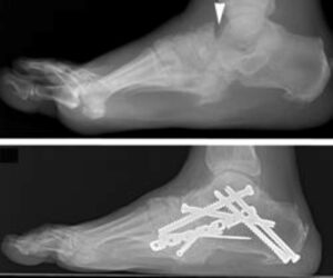
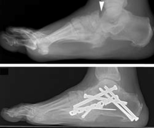
(Top) In this X-ray taken from the side, the patient has unstable Charcot of the back of the foot (hindfoot). The dislocation of the joints is seen where the two bones in the back of the foot do not line up (arrowhead). (Bottom) A complex realignment and fusion was performed to prevent the patient from developing a prominence and ulceration.


Amputation

In severe cases of Charcot arthropathy, where there is significant deformity, deep bone infection, or a loss of blood supply to the foot, salvaging the foot may not be possible. In these situations, an amputation above the affected area may be recommended. Following amputation, patients are typically fitted with a prosthetic device to restore mobility and enhance quality of life.

Surgical Outcomes

Surgery for Charcot foot carries higher risks compared to standard foot and ankle procedures due to underlying diabetes and the extent of deformities. Potential complications include wound healing issues, infections, and, in severe cases, the need for amputation. To minimize these risks, patients should:

  1. Control Blood Sugar: Complication rates are closely linked to hemoglobin A1C levels; maintaining optimal glucose control is critical.
  2. Adhere to Non-Weightbearing Instructions: Patients must avoid placing weight on the foot until cleared by their doctor to reduce the risk of complications.

Each surgical case requires a careful evaluation of risks and benefits. Success and complication rates vary depending on the severity of the condition and the specific procedures performed.

Although surgical risks are significant, the consequences of leaving Charcot foot untreated are often more severe. For example, Charcot patients with an ulcer face a 50% chance of amputation. Therefore, correcting deformities through surgery to prevent ulcers may outweigh the potential surgical risks.

  • Pre-Ulcer Surgery Success Rates: When surgery is performed before ulcers develop, success rates are typically excellent, exceeding 80%.
  • Post-Ulcer Challenges: Surgery after ulcer formation becomes more complex due to the increased risk of infection, significantly lowering the likelihood of a successful outcome.

Post-Surgical Recovery

After surgery, patients usually require a strict non-weightbearing period lasting at least three months. Failure to follow postoperative instructions, including early weightbearing, can lead to serious complications such as:

  • Poor wound healing
  • New injuries
  • Recurrence or worsening of deformities

Adhering to the doctor’s guidelines during recovery is essential to achieve the best outcomes and avoid further complications.


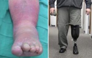
This patient developed an infection of the bone 9 months after an ankle joint fusion to reconstruct a Charcot deformity. After attempts to cure the infection were unsuccessful, it was necessary to amputate his foot and ankle. He returned to walking pain-free with a prosthesis.


Prevention: The Best Approach to Diabetic Foot Care

Preventing diabetic foot problems is the most effective strategy for maintaining foot health. Key preventive measures include:

  • Tight Blood Sugar Control: Maintaining optimal glucose levels helps reduce the risk of complications.
  • Daily Foot Inspections: Regular checks help identify early signs of wounds, ulcers, or other abnormalities.
  • Proper Footwear: Strong, supportive, and full-coverage diabetic shoes are crucial to protecting feet from injuries and pressure points.

For additional guidance, read: Care of the Diabetic Foot for tips on managing foot health.

Early Action is Critical

If you notice swelling, redness, warmth, or any wounds on your foot, consult your doctor immediately. Early diagnosis and treatment of Charcot arthropathy lead to significantly better outcomes.

Managing Charcot Foot

Once diagnosed with Charcot arthropathy, the primary treatment goal is to stabilize the bones in a position that minimizes the risk of ulcers. This can sometimes be achieved with nonsurgical methods, but surgery is often necessary to prevent severe deformities and ulcer formation. Since ulcers increase the risk of amputation by 40%, preventing them is critical.

Tips for Successful Treatment and Long-Term Care

To achieve the best possible outcome:

  1. Follow Medical Instructions: Adhere to your doctor’s recommendations regarding when it is safe to bear weight on the affected foot.
  2. Commit to Lifelong Care: Inspect your feet daily for any changes, and maintain strict blood sugar control to minimize future risks.

Early recognition and consistent management of Charcot foot are essential to avoiding long-term complications and maintaining mobility.

Scroll to Top