Anatomy
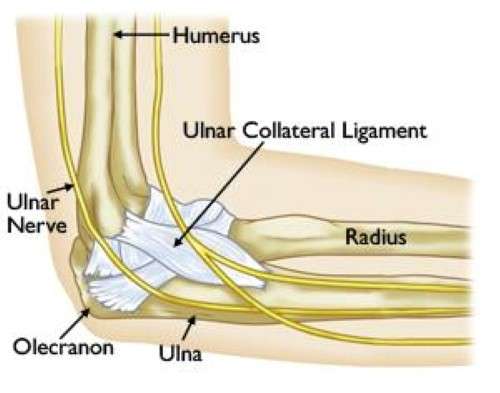
The normal anatomy of the elbow joint shown from the side closest to the body. The bones, major nerves, and ligaments are highlighted.
The elbow joint is where three bones in the arm meet: the upper arm bone (humerus) and the two bones in the forearm (radius and ulna). It is a combination hinge and pivot joint. The hinge part of the joint lets the arm bend and straighten; the pivot part lets the lower arm twist and rotate.
At the upper end of the ulna is the olecranon, the bony point of the elbow that can easily be felt beneath the skin.
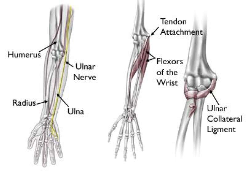
On the inner and outer sides of the elbow, thick ligaments (collateral ligaments) hold the elbow joint together and prevent dislocation. The ligament on the inner part the elbow (closer to the body) is the ulnar collateral ligament. It runs from the inner side of the humerus to the inner side of the ulna and must withstand extreme stresses as it stabilizes the elbow during overhand throwing.
Several muscles, nerves, and tendons (connective tissues between muscles and bones) cross at the elbow. The flexor/pronator muscles of the forearm and wrist begin at the elbow and are also important stabilizers of the elbow during throwing.
The ulnar nerve crosses the elbow joint right behind the bony prominence on the inner aspect of the elbow. It controls the muscles of the hand and provides sensation to the small and ring fingers.
Reproduced and adapted with permission from J Bernstein, ed: Musculoskeletal Medicine. Rosemont, IL, American Academy of Orthopaedic Surgeons, 2003.
Description
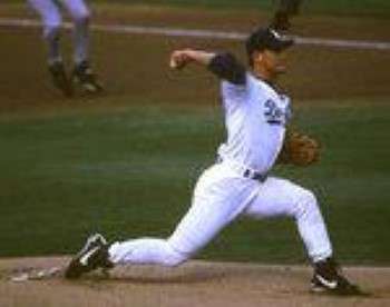
When athletes throw repeatedly at high speed, the repetitive stresses can lead to a wide range of overuse injuries. Problems most often occur at the inside of the elbow because considerable force is concentrated over the inner elbow during throwing.
Reproduced with permission from Ahmad CS, ElAttrache NS: Elbow valgus instability in throwing athletes. Orthopaedic Knowledge Online Journal 2004. Accessed December 2012.
Common Throwing Injuries of the Elbow
Flexor Tendinitis
Repetitive throwing can irritate and inflame the flexor/pronator tendons where they attach to the humerus bone on the inner side of the elbow. Athletes will have pain on the inside of the elbow when throwing, and if the tendinitis is severe, they will also experience pain during rest.
Ulnar Collateral Ligament Injury
The ulnar collateral ligament (UCL) is the most commonly injured ligament in throwers. Injuries of the UCL can range from minor damage and inflammation to a complete tear of the ligament. Athletes will have pain on the inside of the elbow, and frequently notice decreased throwing velocity.
Valgus Extension Overload
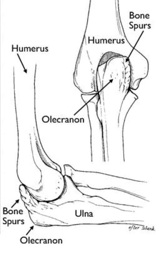
During the throwing motion, the olecranon and humerus bones are twisted and forced against each other. Over time, this can lead to valgus extension overload (VEO), a condition in which the protective cartilage on the olecranon is worn away and abnormal overgrowth of bone called bone spurs, or osteophytes, develop. Athletes with VEO experience swelling and pain at the site of maximum contact between the bones in the back part of the elbow.
Reproduced with permission from Miller CD, Savoie FH III: Valgus extension injuries of the elbow in the throwing athlete. J Am Acad Orthop Surg 1994; 2:261-269.
Olecranon Stress Fracture
Ulnar Neuritis
When the elbow is bent, the ulnar nerve stretches around the bony bump at the inner end of the humerus. In throwing athletes, the ulnar nerve is stretched repeatedly, and can even slip out of place, causing painful snapping. This stretching or snapping leads to irritation of the nerve, a condition called ulnar neuritis.
Throwers with ulnar neuritis will notice pain that resembles electric shocks starting at the inner elbow (often called the “funny bone”) and running along the nerve as it passes into the forearm. They may have numbness, tingling, or pain in the small and ring fingers during or immediately after throwing, and these symptoms may also persist during periods of rest.
Ulnar neuritis can also occur in non-throwers, who frequently notice these same symptoms when first waking up in the morning, or when holding the elbow in a bent position for prolonged periods.
Cause
Elbow injuries in throwers are usually the result of overuse and repetitive high stresses. In many cases, pain will resolve when the athlete stops throwing. It is uncommon for many of these injuries to occur in non-throwers.
In baseball pitchers, rate of injury is highly related to the number of pitches thrown, the number of innings pitched, and the number of months spent pitching each year. Taller and heavier pitchers, pitchers who throw with higher velocity, and those who participate in showcases are also at higher risk of injury. Pitchers who throw with arm pain or while fatigued have the highest rate of injury.
Symptoms
Most elbow injuries initially cause pain during or after throwing. They will often limit the ability to throw or decrease throwing velocity. the athletes or coaches may also notice that pitches are starting to sail high. In the case of ulnar neuritis, the athlete will frequently experience numbness and tingling of the elbow, forearm, or hand as described above.
Doctor Examination
Reproduced with permission from JF Sarwark, ed: Essentials of Musculoskeletal Care, ed 4. Rosemont, IL, American Academy of Orthopaedic Surgeons, 2010.
During the physical examination, the doctor will check the range of motion, strength, and stability of the elbow. They may also evaluate the athlete’s shoulder.
The doctor will also assess muscle bulk and appearance, and will compare the injured elbow with the opposite side. In some cases, they will assess sensation and individual muscle strength.
The doctor will ask the athlete to identify the area of greatest pain, and will frequently use direct pressure over several distinct areas to try to pinpoint the exact location of the pain.
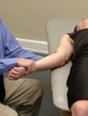
To recreate the stresses placed on the elbow during throwing, the doctor will perform the valgus stress test. During this test, the doctor holds the arm still and applies pressure against the side of the elbow. If the elbow is loose or if this test causes pain, it is considered a positive test. Other specialized physical examination maneuvers may be necessary, as well.
The results of these tests help the doctor decide if additional testing or imaging of the elbow is necessary.
Imaging Tests
X-rays. X-rays provide clear pictures of dense structures, like bone. They will often show stress fractures, bone spurs, and other abnormalities.
Computed tomography (CT) scans. CT scans provide a three-dimensional image of bony structures and can be very helpful in defining bone spurs or other bony disorders that may limit motion or cause pain. These scans are not typically used to help diagnose problems in throwers’ elbows.
Magnetic resonance imaging (MRI) scans. MRI scans provide an excellent view of the soft tissues of the elbow and can help the doctor distinguish between ligament and tendon disorders that often cause the same symptoms and physical examination findings. MRI scans can also help determine the severity of an injury, such as whether a ligament is mildly damaged or completely torn. MRI is also useful in identifying a stress fracture that is not visible in an X-ray image. In some cases, the doctor may order an arthrogram, in which dye is injected into the elbow joint, and an MRI scan is then taken. This test can assess for ligament tears.
Treatment
Nonsurgical Treatment
In most cases, treatment for throwing injuries in the elbow begins with a short period of rest.
Additional treatment options may include:
Physical therapy. Specific exercises can restore flexibility and strength. A rehabilitation program directed by the doctor or a physical therapist will include a gradual return to throwing.
Change of position. Throwing mechanics can be evaluated in order to correct body positioning that puts excessive stress on the elbow.
Although a change of position or even a change in sport can eliminate repetitive stresses on the elbow and provide lasting relief, this is often undesirable, especially in high level athletes.
Anti-inflammatory medications. Drugs like ibuprofen and naproxen reduce pain and swelling and can be provided in prescription-strength form.
If symptoms persist, the athlete may need a prolonged period of rest.
Injections. In some cases, an injection of platelet-rich plasma (PRP) can be beneficial in patients with partial tearing of the UCL. There is growing evidence in the literature to support use of PRP, which involves using the patient’s own platelets to stimulate healing. For this procedure, a small amount of blood is drawn from the patient. The platelets are then separated from other blood cells using a centrifuge and injected into the area of the injury.
Surgical Treatment
If painful symptoms are not relieved by nonsurgical methods, and the athlete desires to continue throwing, surgical treatment may be considered.
Arthroscopy. Bone spurs on the olecranon and any loose fragments of bone or cartilage within the elbow joint can be removed arthroscopically.
During arthroscopy, the surgeon inserts a small camera, called an arthroscope, into the elbow joint. The camera displays pictures on a television screen, and the surgeon uses these images to guide miniature surgical instruments.
Because the arthroscope and surgical instruments are thin, the surgeon can use very small incisions, rather than the larger incision needed for standard, open surgery.
UCL reconstruction. Athletes who have an unstable or torn UCL, and who do not respond to nonsurgical treatment, are candidates for surgical ligament reconstruction.
Most ligament tears cannot be sutured (stitched) back together. To surgically repair the UCL and restore elbow strength and stability, the ligament must be reconstructed. During the procedure, the doctor replaces the torn ligament with a tissue graft. This graft acts as a scaffolding for a new ligament to grow on. In most cases of UCL injury, the ligament can be reconstructed using one of the patient’s own tendons.
This surgical procedure is often referred to as “Tommy John surgery,” named after the former major league pitcher who underwent the first successful UCL reconstruction in 1974. Today, UCL reconstruction has become a common procedure. Though return to play is not guaranteed, the procedure has helped professional and college athletes continue to compete in a range of sports.
In some cases, if the ligament is in good condition but is torn at the bony attachment, it can be reattached to the arm, eliminating the need for a graft. sometimes, the ligament is reinforced with a high-strength suture to add to the strength of the construct and potentially allow for a quicker return to play.
Ulnar nerve anterior transposition. In cases of ulnar neuritis, the nerve can be moved to the front of the elbow to prevent stretching or snapping. This is called an anterior transposition of the ulnar nerve.
Recovery
If nonsurgical treatment is effective, the athlete can often return to throwing in 6 to 9 weeks.
If surgery is required, however, recovery may take much longer, depending upon the procedure performed. For example, it may take the athlete 6 to 9 months or more to return to competitive throwing after UCL reconstruction.
Prevention
Recent research has focused on identifying risk factors for elbow injury and strategies for injury prevention.
Proper conditioning, technique, and recovery time can help to prevent throwing injuries in the elbow.
In the case of younger athletes, pitching guidelines — including pitch count limits and required rest recommendations — have been developed to protect children from injury.

