Understanding Patellofemoral Replacement
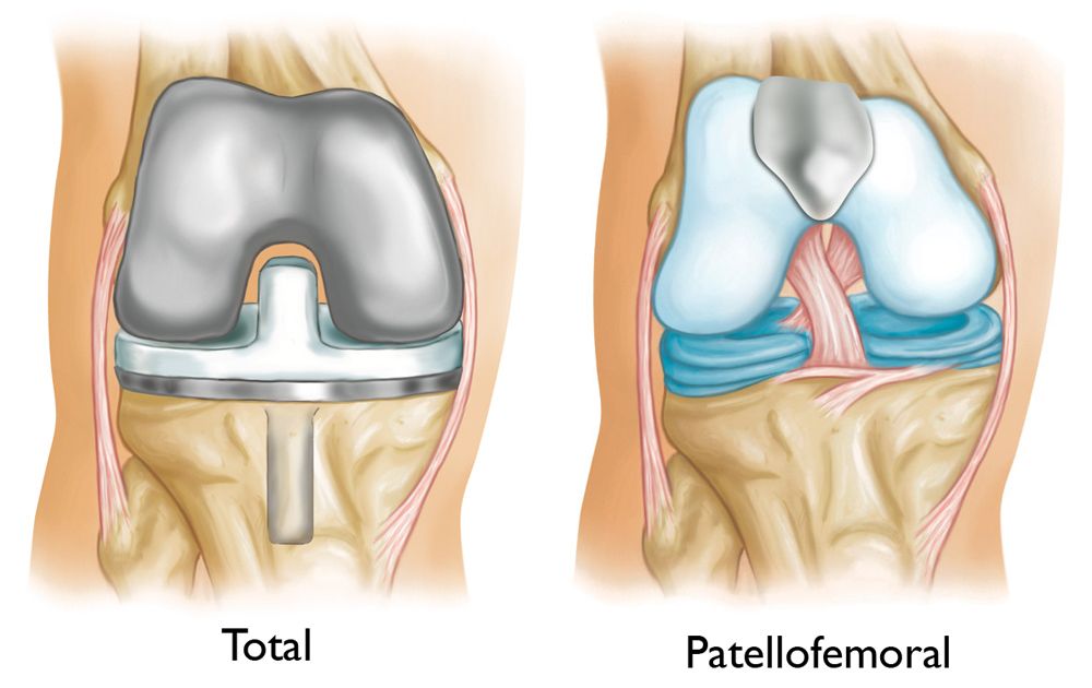
In a total knee replacement, all three compartments of the knee with damaged bone and cartilage are resurfaced using metal and plastic components.
Patellofemoral replacement, on the other hand, is a type of partial knee replacement that focuses on resurfacing only a specific portion of the knee. It is a suitable option for patients whose bone and cartilage damage is confined to the underside of the patella (kneecap) and the trochlear groove in the femur (thighbone) where the kneecap rests. This condition is known as patellofemoral arthritis.
Compared to total knee replacement, patellofemoral replacement is performed through a smaller incision, causing less disruption to the surrounding soft tissues. As a result, many patients experience a faster recovery and return to daily activities sooner than those undergoing total knee replacement.
There are several treatment options available for knee osteoarthritis. Your doctor will discuss the most appropriate options to effectively address your specific symptoms and improve your quality of life.
Anatomy of the Knee
The knee joint is divided into three major compartments:
- Medial Compartment: The inner portion of the knee.
- Lateral Compartment: The outer portion of the knee.
- Patellofemoral Compartment: The front portion of the knee, located between the patella (kneecap) and the femur (thighbone).
Within the patellofemoral compartment, the patella rests in a groove at the top of the femur known as the trochlear groove. When you bend or straighten your knee, the patella glides smoothly back and forth within this groove.
The ends of the femur, the trochlear groove, and the underside of the patella are covered by articular cartilage, a smooth and protective substance that allows the bones to glide seamlessly against each other during movement.
This structure plays a vital role in maintaining knee function and mobility, particularly in activities involving bending, straightening, or weight-bearing.
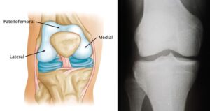
Left) A normal knee joint. The medial, lateral and patellofemoral compartments are shown.
(Right) An X-ray of a normal knee showing healthy space between the bones.
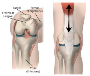
(Right) As you bend and straighten your knee, the patella slides up and down within the groove.
Description of Patellofemoral Replacement
In knee osteoarthritis, the protective cartilage covering the bones of the knee gradually wears away. As the cartilage deteriorates, it becomes frayed, exposing the underlying bone. The movement of bones over this rough, unprotected surface causes significant pain. Osteoarthritis can affect the entire knee joint or be limited to a single area.
When advanced osteoarthritis is confined to the patellofemoral compartment (the area between the kneecap and the femur), it may be treated with a patellofemoral replacement. During this procedure:
- The underside of the kneecap is resurfaced with a plastic implant.
- The trochlear groove of the femur is resurfaced with a metal implant.
The healthy cartilage, bone, and all supporting ligaments in the rest of the knee are carefully preserved, ensuring that the natural knee structure remains intact while alleviating pain and restoring function.
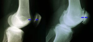
(Left) This X-ray shows a normal knee from the side. The arrows point to the normal amount of space between the bones.
(Right) This X-ray shows narrowed joint space and bone rubbing on bone due to arthritis.
Advantages of Patellofemoral Replacement
Patellofemoral replacement offers several potential benefits compared to total knee replacement, particularly for patients with localized knee arthritis. These advantages include:
- Less Blood Loss: Reduced surgical intervention leads to minimal blood loss.
- Smaller Incision and Less Trauma: The procedure involves a smaller surgical incision and causes less damage to surrounding tissues.
- Reduced Pain and Swelling: Patients typically experience less post-surgical discomfort and swelling.
- Faster Recovery: A quicker return to normal activities compared to total knee replacement.
- Lower Risk of Complications: Fewer risks associated with a less invasive procedure.
- Improved Knee Function and Activity: Many patients achieve increased mobility and better knee performance.
Additionally, because the healthy bone, cartilage, and ligaments in the rest of the knee are preserved, many patients report that a patellofemoral replacement provides a more natural feel compared to a total knee replacement.
Disadvantages of Patellofemoral Replacement
The primary drawback of patellofemoral replacement compared to total knee replacement is the potential need for additional surgery in the future. If arthritis progresses and develops in the remaining compartments of the knee that were not resurfaced, a total knee replacement may eventually become necessary to address the new areas of damage.
Candidates for Patellofemoral Replacement
If your osteoarthritis has progressed and nonsurgical treatments no longer provide relief, your doctor may recommend knee replacement surgery. However, patellofemoral replacement is only suitable for specific patients, and careful evaluation is essential.
To be considered a candidate, your arthritis must be limited to the patellofemoral compartment of the knee.
Conditions That May Exclude You as a Candidate
You may not be eligible for the procedure if you have:
- Knee stiffness
- Ligament damage
- Poor patellar tracking (improper kneecap movement)
- Significant leg deformity
- Inflammatory arthritis (e.g., rheumatoid arthritis)
- Crystalline arthritis (e.g., gout)
- Morbid obesity
Your doctor will assess your overall knee condition and health to determine if patellofemoral replacement is the most appropriate treatment for you.
Orthopaedic Evaluation for Patellofemoral Replacement
A comprehensive evaluation by an orthopedic surgeon is essential to determine if you are a suitable candidate for patellofemoral replacement. This process involves reviewing your medical history, performing a physical examination, and conducting imaging tests.
Medical History
Your doctor will ask detailed questions about your general health, knee pain, and functional limitations.
- Pain Location: Identifying the exact location of your pain is critical. Candidates for patellofemoral replacement typically experience pain behind the kneecap or in the anterior knee. This pain is often triggered by activities that place stress on the kneecap, such as:
- Climbing stairs
- Sitting with the knee bent
- Rising from a chair
Physical Examination
Your doctor will perform a thorough physical exam to pinpoint the source of your pain and assess the knee’s overall condition. This includes:
- Inspecting the Knee: Evaluating the alignment of the joint.
- Palpating the Knee: Pressing on specific areas to reproduce your pain.
- Range of Motion Testing: Checking for stiffness or issues with patellar tracking (abnormal movement of the kneecap during leg bending or straightening).
- Stability Assessment: Evaluating the strength and integrity of the ligaments surrounding the knee.
Imaging Tests
- X-rays: X-rays provide clear images of the bones. Your doctor will order X-rays from multiple angles to confirm that arthritis is confined to the patellofemoral compartment and to evaluate knee alignment.
- MRI Scans: MRI scans offer detailed images of soft tissues. Your doctor may request an MRI to assess the condition of the cartilage and soft tissues in your knee more precisely.
These evaluations collectively help your orthopedic surgeon determine whether patellofemoral replacement is the right treatment for your specific condition.
Your Surgery
Patients undergoing patellofemoral replacement typically experience faster recovery than those undergoing total knee replacement. As a result, the procedure is often performed on an outpatient basis. During your initial consultation, your doctor will evaluate whether you are suitable for outpatient surgery or require a short hospital stay.
Before Surgery
On the day of your surgery:
- Your surgeon will verify the surgical site by marking the correct knee with a marker.
- A doctor from the anesthesia team will discuss your options, which may include:
- General anesthesia: You are completely asleep.
- Spinal anesthesia: You remain awake, but your body is numb from the waist down.
These options should also have been discussed with your surgeon during preoperative visits.
Surgical Procedure
1. Joint Inspection
The surgeon begins by making an incision at the front of your knee and inspecting all three compartments of the joint to confirm that the damaged cartilage is limited to the patellofemoral compartment. They will also check the integrity of your ligaments.
- If damage extends beyond the patellofemoral compartment, a total knee replacement may be performed instead. This contingency plan will be discussed with you before surgery to ensure you agree with this approach if needed.
2. Patellofemoral Replacement
The procedure involves two main steps:
- Preparing the Bone: Special tools are used to remove damaged cartilage and a small amount of bone from the patellofemoral compartment.
- Positioning the Implants:
- A plastic button is used to resurface the underside of the patella (kneecap).
- A metal component is used to resurface the trochlear groove of the femur.
- Both parts are securely attached to the bone with surgical cement to ensure stability and proper alignment.
By preserving healthy structures and targeting only the damaged area, patellofemoral replacement aims to restore knee function with minimal impact on surrounding tissues.
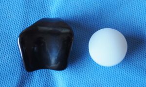
Patellofemoral replacement implants. The metal femoral implant (left) resurfaces the trochlear groove of the femur. The round plastic patellar implant (right) attaches to the underside of the kneecap.

(Left) This X-ray is taken from above the knee. The patella and the trochlear groove of the femur have become deformed due to osteoarthritis. There is now bone rubbing on bone. (Right) The same knee after patellofemoral replacement. The patellar implant on the underside of the kneecap does not show in an X-ray.
After Surgery
Following the procedure, you will be moved to the recovery room, where medical staff will closely monitor you as you awaken from the anesthesia. Once your condition stabilizes:
- If your surgery was performed on an outpatient basis, you will be discharged the same day with post-operative instructions.
- If a hospital stay is required, you will be transferred to your hospital room for further observation and recovery.
Your medical team will ensure that you are comfortable and provide guidance for the next steps in your recovery process.
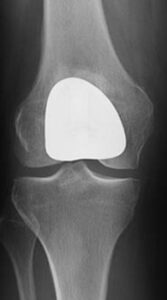
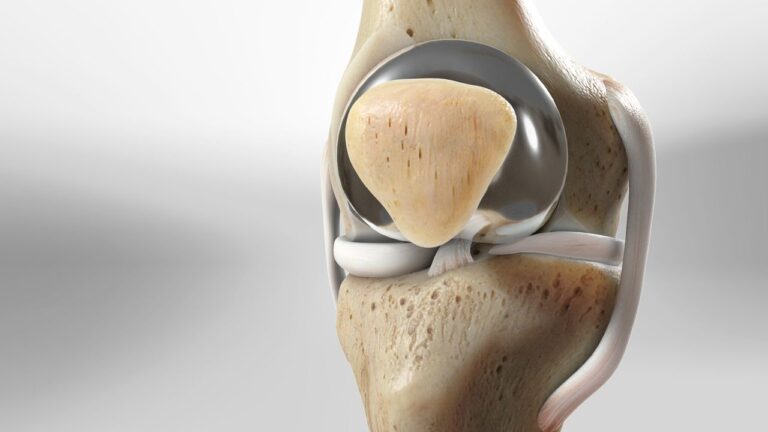
Front view of a knee after patellofemoral replacement.
Recovery After Patellofemoral Replacement
Pain Management
Post-surgery, you may experience some pain, which can be effectively managed with various medications, including:
- Opioids: These narcotics provide strong pain relief but must be used strictly as directed due to their addictive potential. Stop taking them as soon as your pain improves.
- Nonsteroidal Anti-Inflammatory Drugs (NSAIDs): Help reduce pain and inflammation.
- Local Anesthetics: May be used to provide localized relief.
Effective pain management not only improves comfort but also supports faster healing and recovery.
Weight-Bearing
You will begin placing weight on your knee immediately after surgery. However, you may need assistance from a walker, cane, or crutches for a few days as your knee adjusts to the new implants and heals.
Rehabilitation Exercises
A physical therapist will guide you through specific exercises designed to:
- Strengthen your quadriceps muscles.
- Maintain and improve the range of motion in your knee.
Performing these exercises consistently and as directed is crucial for achieving the best possible outcome.
Follow-Up Visits
Regular follow-up appointments with your orthopedic surgeon are essential to monitor your progress. These visits will ensure proper healing and alignment of the implants, allowing adjustments to your recovery plan as needed.

