The femur, commonly known as the thighbone, is the body’s largest and strongest bone. Its robust structure typically requires a significant amount of force to fracture. In children, femur fractures often occur due to sudden and intense impacts, such as those experienced in accidents or falls.
Anatomy of the Femur
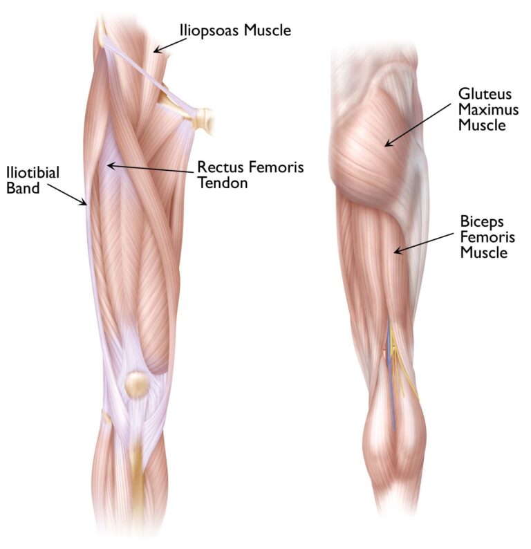
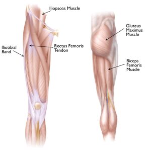
The femur, or thighbone, runs from the hip joint to the knee and serves as the body’s primary load-bearing bone. The long, straight section of this bone is referred to as the femoral shaft. When a fracture occurs anywhere along this portion, it is classified as a femoral shaft fracture.
Causes of Femur Fractures in Children
In infants under one year of age, child abuse is the most frequent cause of femur fractures. While child abuse also accounts for many femur fractures in children aged 1 to 4, its occurrence decreases significantly as children grow older.
For adolescents, motor vehicle accidents—whether as passengers, bicyclists, or pedestrians—are the leading cause of femoral shaft fractures. Other high-risk activities for pediatric femur fractures include:
- Hard falls on playground equipment
- High-impact collisions during contact sports
- Motor vehicle accidents
- Abuse in non-walking infants
Types and Classification of Femur Fractures
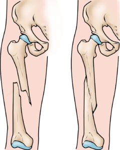
Femur fractures can vary significantly in their nature. The bone fragments may remain properly aligned or become misaligned (displaced). Fractures may also be closed, where the skin remains intact, or open, where the broken bone pierces through the skin—though open fractures are uncommon.
These fractures are categorized based on:
- Location: The fracture may occur at the top, middle, or bottom of the femur.
- Shape: Breaks can take various forms, such as transverse (straight across), oblique (angled), or spiral (twisted).
- Alignment: Fractures may result in angled or displaced bone ends.
- Fragmentation: Bones that shatter into three or more pieces are termed comminuted fractures.
By understanding these classifications, healthcare providers can determine the best treatment approach tailored to the type and severity of the fracture.
 Types of femur fractures. (Left) An oblique, displaced fracture of the femur shaft. (Right) A comminuted fracture of the femoral shaft.
Symptoms of a Femur Fracture
A femur fracture is a significant injury, often with unmistakable signs. Common symptoms indicating a potential thighbone fracture in children include:
- Severe Pain: Intense discomfort in the thigh region.
- Swelling or Deformity: Noticeable swelling or an abnormal shape in the thigh.
- Inability to Stand or Walk: Your child may be unable to bear weight, stand, or walk.
- Limited Range of Motion: Pain may restrict movement in the hip or knee.
If you observe any of these symptoms, it’s crucial to seek immediate medical attention by taking your child to the emergency room. Early diagnosis and treatment are essential for proper healing and recovery.
Doctor Examination: Key Steps for Diagnosing a Femur Fracture
When evaluating a suspected femur fracture, it is crucial for the doctor to understand the details surrounding the injury. Be prepared to share if your child experienced any pre-existing conditions, illnesses, or other trauma before the fracture occurred.
During the examination:
- Pain Management: The doctor will administer pain relief medication to ensure your child is comfortable.
- Thorough Assessment: The entire leg, including the hip and knee, will be carefully examined. Since femur fractures can be associated with other severe injuries, a comprehensive evaluation is essential.
Imaging Tests for Accurate Diagnosis
- X-Rays: X-rays are a primary diagnostic tool used to locate the fracture, determine its type, and assess bone alignment. This imaging also helps the doctor evaluate the growth plate near the end of the femur, which is critical for bone development.
- Growth Plate Monitoring: If the growth plate is affected, surgical intervention may be necessary to restore its function. Regular x-rays over several months may be required to monitor healing and ensure proper bone growth.
Treatment Options for Femur Fractures
Treatment plans depend on several factors, including the child’s age, weight, type of fracture, cause of injury, and whether the skin was broken by the bone. The primary goal is to realign the bone fragments and stabilize them for optimal healing.
Nonsurgical Treatment
- Closed Reduction: In certain cases, the doctor can manually realign the broken bones without surgery.
- Pavlik Harness: For infants under six months old, a Pavlik harness can immobilize the fractured bone, allowing it to heal effectively.
- Spica Casting:
For children aged 7 months to 5 years, a spica cast is commonly used to maintain proper alignment during the healing process.- Design: A spica cast typically extends from the chest down to the fractured leg. It may also cover the uninjured leg partially or fully, depending on the fracture’s specifics.
- Customization: The type of spica cast is chosen based on the fracture’s characteristics and what will provide the most effective stabilization.
By following these diagnostic and treatment protocols, doctors aim to achieve successful recovery while ensuring the bone continues to grow and function properly.
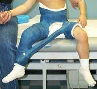 A young child in a hip spica cast to immobilize a femoral shaft fracture.
Spica Casting Procedure for Femur Fractures
To perform a closed reduction, your doctor will sedate your child to ensure they remain comfortable during the procedure. Following the realignment, a spica cast will be applied either immediately or within 24 hours of hospitalization. This cast helps maintain the correct position of the fractured bone pieces during the healing process.
When a bone is fractured and displaced, the broken ends often overlap, leading to a temporary shortening of the bone’s normal length. However, because children’s bones grow and remodel quickly, perfect alignment is not always necessary. While in the spica cast, the bone will naturally heal and reshape toward a normal configuration.
For optimal outcomes:
- Minimal Overlap: The broken bone ends should not overlap more than 2 centimeters while in the cast.
- Healing and Growth: The trauma from the fracture may temporarily accelerate femur growth, helping correct any mild shortening caused by the overlap. Over time, this process ensures the bone regains its normal length and function.
This approach leverages a child’s natural growth and healing capabilities to achieve effective recovery with minimal intervention.
A femur fracture before and immediately after treatment with a spica cast. The femur will remodel over time so that it appears normal.
Traction for Femur Fractures
If the bone ends are shortened by more than 3 centimeters or if the alignment is too crooked despite the use of a cast, traction may be recommended. This technique involves a steady, gentle pulling force applied to the leg to properly realign the bones before other treatments are initiated.
Surgical Treatment for Femur Fractures
For femur fractures with significant displacement—specifically those with more than 3 centimeters of shortening—surgical intervention is often necessary to correct the alignment and stabilize the bone. In complex cases, the bone may need to be surgically realigned and supported with an implant to ensure proper healing.
Surgical management of pediatric femur fractures has become increasingly common due to its recognized advantages, including:
- Faster recovery and rehabilitation
- Earlier mobilization of the child
- Reduced hospital stays
Flexible Intramedullary Nails for Stabilization
In children aged 6 to 10 years, flexible intramedullary nails have become a preferred method for stabilizing femur fractures. These nails are placed inside the bone to maintain alignment during healing. Over the past decade, this approach has gained widespread acceptance due to its effectiveness and minimal invasiveness, allowing children to recover more quickly and resume normal activities sooner.
By employing tailored treatments like traction or surgery, doctors can ensure optimal outcomes based on the severity and specifics of the fracture.
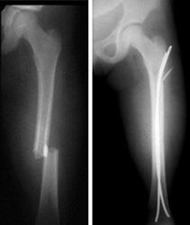
(Left) Preoperative x-ray of a child with a fracture through the middle of the shaft of the left femur. (Right) Postoperative x-ray of the same child shows that the fracture was treated with internal flexible nailing to restore stability and allow early mobilization.
Alternative Surgical Treatments for Complex Femur Fractures
In cases where the femur fracture involves multiple bone fragments, flexible intramedullary nails may not be effective. For such complex injuries, other surgical options can achieve successful outcomes, including:
- Plates and Screws: A plate with screws may be used to “bridge” the fractured segments, providing stable alignment while the bone heals.
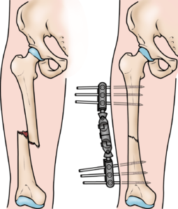
- External Fixator: A stabilizing frame outside the body can be used, particularly in cases involving extensive open injuries to the skin and surrounding muscles. This method protects the area while promoting healing.
- Prolonged Traction with Temporary Pins: When other treatments are not viable, traction using a temporary pin inserted into the bone can realign the fragments over time.
Each of these methods is carefully chosen based on the fracture’s complexity and the child’s overall health, ensuring the best possible recovery while minimizing complications.
External fixation is often used to hold the bones together when the skin and muscles have been injured.
Treatment Options for Older Children and Adolescents
As children approach their teenage years (11 years and older, up to skeletal maturity), the treatment of femur fractures often involves one of two options:
- Flexible Intramedullary Nails: These nails are suitable for many fractures and provide stability while allowing the child to begin walking soon after the procedure.
- Rigid Locked Intramedullary Nails: This option is particularly beneficial for fractures that are unstable or more complex. The rigid nail provides enhanced support and ensures proper alignment during healing.
Both methods are designed to enable early mobility, which is crucial for faster recovery and reduced long-term complications. The choice between flexible and rigid nails depends on the fracture’s characteristics and the child’s overall condition.
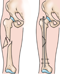 A rigid, locked intramedullary nail is often used for femur fractures in adolescents who are nearly full grown.
Long-Term Outcomes for Pediatric Femur Fractures
Most children who experience a femur fracture recover fully, regaining normal leg function with equal leg lengths. In some cases, intramedullary nails used during treatment may need to be removed after healing if they cause irritation to the surrounding skin or tissues.
Occasionally, additional treatment may be necessary, either shortly after the initial recovery or later in life, to address specific complications, such as:
- Significant differences in leg length
- Unacceptable angulation of the healed bone
- Abnormal rotation of the bone after healing
- Persistent bone infection
- Rare instances of nonunion, where the fracture does not heal properly
These issues are typically manageable with further medical intervention, ensuring that children can achieve long-term functional and structural recovery.

